Calculate Total Magnification Of A Microscope
Muz Play
Apr 06, 2025 · 5 min read

Table of Contents
Calculating Total Magnification of a Microscope: A Comprehensive Guide
Understanding the total magnification of your microscope is crucial for any microscopy work, whether you're a seasoned researcher or a curious student. Total magnification determines the size of the image you see, directly impacting your ability to resolve fine details and make accurate observations. This comprehensive guide will walk you through the process of calculating total magnification, exploring the factors involved, and providing practical examples to solidify your understanding.
Understanding Magnification: The Basics
Before diving into calculations, let's establish a clear understanding of magnification. Magnification is simply the process of enlarging an image. Microscopes achieve this using a combination of lenses that bend light to create a magnified virtual image of the specimen. This process is crucial for visualizing structures too small to be seen with the naked eye.
Types of Lenses: The Key Players
A typical compound light microscope uses two main types of lenses:
-
Objective Lenses: These are the lenses closest to the specimen. They provide the initial magnification of the image. A standard microscope often includes multiple objective lenses with different magnifications, commonly 4x, 10x, 40x, and 100x. The 100x objective typically requires immersion oil for optimal performance.
-
Eyepiece (Ocular) Lens: This is the lens you look through. It further magnifies the image produced by the objective lens. Most eyepieces have a magnification of 10x.
Calculating Total Magnification: The Formula
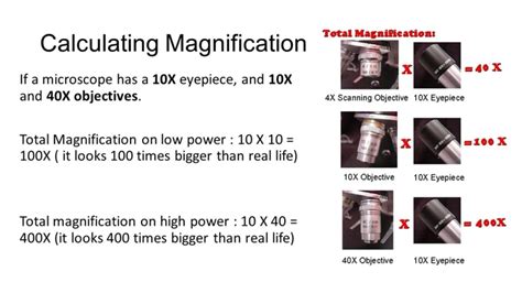
The total magnification of a microscope is the product of the objective lens magnification and the eyepiece lens magnification. This can be expressed by the following simple formula:
Total Magnification = Objective Lens Magnification × Eyepiece Lens Magnification
This formula is fundamental to understanding the magnification you're achieving. Let's illustrate this with some examples.
Practical Examples: Calculating Total Magnification
Let's consider several scenarios to demonstrate how to apply the total magnification formula:
Scenario 1: Standard Observation
You're using a 10x objective lens and a 10x eyepiece. Applying the formula:
Total Magnification = 10x × 10x = 100x
This means you're viewing the specimen at a magnification of 100 times its actual size.
Scenario 2: High-Magnification Imaging
You switch to a 40x objective lens while keeping the same 10x eyepiece.
Total Magnification = 40x × 10x = 400x
Now, you're observing the specimen at 400 times its actual size, revealing much finer details.
Scenario 3: Immersion Oil Objective
You utilize the 100x oil immersion objective lens with the 10x eyepiece.
Total Magnification = 100x × 10x = 1000x
This high magnification allows for the visualization of extremely small structures, but requires careful technique and the use of immersion oil to maintain image clarity.
Scenario 4: Different Eyepiece Magnification
Suppose your microscope has a 15x eyepiece. Using the 40x objective lens:
Total Magnification = 40x × 15x = 600x
This demonstrates how a change in eyepiece magnification directly affects the total magnification.
Beyond the Formula: Factors Affecting Image Quality
While the formula for total magnification is straightforward, obtaining a clear and sharp image involves several other factors:
Resolution: Seeing the Fine Details
Resolution refers to the ability of the microscope to distinguish between two closely spaced points. High total magnification doesn't automatically guarantee high resolution. Numerical aperture (NA) of the objective lens and the wavelength of light used are crucial factors affecting resolution. A higher NA generally provides better resolution.
Numerical Aperture (NA): A Key to Resolution
The numerical aperture (NA) is a measure of a lens's ability to gather light and resolve fine details. A higher NA leads to better resolution and brighter images. You'll find the NA value engraved on the objective lens itself. Understanding NA is vital for interpreting magnification capabilities accurately.
Working Distance: The Space Between Lens and Specimen
The working distance is the distance between the objective lens and the specimen. This distance varies depending on the objective lens. Higher magnification objectives typically have shorter working distances, requiring more precise focusing.
Depth of Field: Focusing on the Z-Axis
Depth of field refers to the vertical range within which the specimen appears in focus. Higher magnification typically results in a shallower depth of field, meaning only a thin section of the specimen will be in sharp focus at any given time.
Illumination: Lighting the Way
Proper illumination is crucial for optimal image quality. Adjusting the light intensity and using appropriate condenser settings are essential for achieving a well-lit and clear image.
Advanced Microscopy Techniques and Magnification
Different microscopy techniques utilize different approaches to achieve magnification and resolution beyond the capabilities of a standard compound light microscope.
Electron Microscopy: Beyond the Limits of Light
Electron microscopy uses beams of electrons instead of light, achieving significantly higher magnification and resolution than light microscopy. Transmission electron microscopy (TEM) and scanning electron microscopy (SEM) are two common types, capable of visualizing structures at the nanometer scale.
Confocal Microscopy: Optical Sectioning for 3D Imaging
Confocal microscopy uses a laser beam to scan the specimen, generating sharp, high-resolution images with improved depth of field. This technique is particularly useful for visualizing three-dimensional structures.
Super-Resolution Microscopy: Breaking the Diffraction Barrier
Super-resolution microscopy techniques bypass the limitations of the diffraction limit of light, allowing for the visualization of structures at a resolution significantly beyond the capabilities of conventional light microscopy. These advanced techniques open up new possibilities for cellular and molecular imaging.
Practical Tips for Accurate Magnification and Image Quality
- Clean Lenses: Ensure your lenses are clean and free from dust or fingerprints, as this can significantly impact image quality.
- Proper Focusing: Take your time to carefully focus the image using the coarse and fine adjustment knobs.
- Optimal Illumination: Adjust the light intensity and condenser settings to achieve even illumination across the field of view.
- Immersion Oil (for 100x objectives): Use immersion oil correctly with the 100x objective lens to avoid damaging the lens and to enhance image clarity.
- Calibration: Regularly calibrate your microscope to ensure accurate magnification readings.
Conclusion: Mastering Total Magnification
Calculating the total magnification of your microscope is a fundamental skill for any microscopy user. By understanding the formula and the factors affecting image quality, you can optimize your microscopy setup to achieve the best possible results. Remember, total magnification is just one aspect of microscopy; resolution, numerical aperture, and proper technique all contribute to the quality of your observations. With practice and a thorough understanding of these concepts, you'll be able to confidently capture clear and informative images of your specimens. This guide provides a strong foundation for your microscopy journey, enabling you to explore the microscopic world with greater precision and insight.
Latest Posts
Latest Posts
-
Testing A Hypothesis About A Population Mean
Apr 06, 2025
-
Can Rate Of Change Be Negative
Apr 06, 2025
-
Energy And Momentum Of Rotating Systems
Apr 06, 2025
-
How Do You Estimate A Decimal
Apr 06, 2025
-
When Is Work Positive Or Negative Thermodynamics
Apr 06, 2025
Related Post
Thank you for visiting our website which covers about Calculate Total Magnification Of A Microscope . We hope the information provided has been useful to you. Feel free to contact us if you have any questions or need further assistance. See you next time and don't miss to bookmark.
