Cardiac Muscles Differ From Skeletal Muscles In That They
Muz Play
Apr 01, 2025 · 7 min read
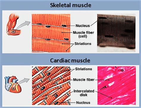
Table of Contents
Cardiac Muscles Differ From Skeletal Muscles in That They… Possess Unique Properties for Life-Sustaining Rhythmic Contractions
Cardiac muscle, the specialized tissue that forms the heart, differs significantly from skeletal muscle, the tissue responsible for voluntary movement. While both are striated muscles, meaning they exhibit a striped pattern under a microscope, their functional properties, cellular structures, and control mechanisms are dramatically different. This fundamental difference reflects their distinct roles in the body: rhythmic, involuntary contractions for the heart versus voluntary, controlled movements for the skeletal system. This article delves deep into the key distinctions between cardiac and skeletal muscle, exploring their structural composition, contraction mechanisms, and regulatory systems.
Structural Differences: A Microscopic Look at Muscle Tissue
At the cellular level, several key distinctions set cardiac muscle apart from skeletal muscle. These differences underpin their contrasting functional capabilities.
1. Cell Structure and Intercalated Discs: The Heart's Unique Connection
Cardiac muscle cells, also known as cardiomyocytes, are branched, shorter, and cylindrical compared to the long, cylindrical fibers of skeletal muscle cells. This branching creates a complex, interconnected network within the heart, vital for coordinated contractions. Perhaps the most defining feature of cardiac muscle is the presence of intercalated discs. These specialized structures are located at the junctions between adjacent cardiomyocytes, appearing as dark lines under a microscope. Intercalated discs are crucial for efficient communication and force transmission between cells.
They contain:
-
Gap junctions: These protein channels directly connect the cytoplasm of adjacent cardiomyocytes, allowing for rapid electrical signal propagation. This rapid spread of electrical impulses ensures synchronized contractions across the entire heart. This is in stark contrast to skeletal muscles, where each fiber is individually innervated.
-
Desmosomes: These strong adhesive junctions provide structural support, anchoring the cells together and preventing them from separating during powerful contractions. The structural integrity provided by desmosomes is essential for the heart's continuous, strenuous workload.
Skeletal muscle cells, on the other hand, are structurally independent units. Although they can be organized into bundles or fascicles, they lack the direct, gap junction-mediated connections found in cardiac muscle. This independent nature allows for fine motor control and individual fiber recruitment in skeletal muscles.
2. Myofibrils and Sarcomeres: The Engines of Contraction
Both cardiac and skeletal muscle contain myofibrils, cylindrical structures composed of repeating units called sarcomeres. Sarcomeres are the basic contractile units of muscle, containing the actin and myosin filaments responsible for generating force. The arrangement of these filaments creates the striated appearance of both muscle types.
However, there are subtle differences in the organization of myofibrils and sarcomeres:
-
Cardiac muscle: Myofibrils in cardiac muscle are often branched and less regularly arranged than in skeletal muscle. This less organized structure is thought to contribute to the cardiac muscle's ability to withstand greater stretching forces.
-
Skeletal muscle: Skeletal muscle myofibrils are highly organized, arranged in parallel arrays running the length of the fiber. This organized structure contributes to the efficiency of force generation in skeletal muscle.
3. Mitochondria: The Powerhouses of Muscle
Cardiac muscle cells are remarkably rich in mitochondria, the cellular powerhouses that generate ATP (adenosine triphosphate), the energy currency of cells. This high mitochondrial density reflects the heart's constant need for energy to maintain its rhythmic contractions throughout life. The heart has an extremely high energy demand, far exceeding that of most other organs. Compromised mitochondrial function can lead to severe cardiac dysfunction.
Skeletal muscle cells also contain mitochondria, but their density is considerably lower than in cardiac muscle, reflecting the intermittent nature of skeletal muscle activity. Skeletal muscle can utilize anaerobic metabolism (producing ATP without oxygen) for short bursts of intense activity, but cardiac muscle relies almost entirely on aerobic respiration (requiring oxygen) for its continuous operation.
Functional Differences: Contraction, Control, and Fatigue
The structural differences between cardiac and skeletal muscle translate into significant functional variations.
1. Contraction Mechanisms: The Role of Calcium
Both cardiac and skeletal muscles utilize the sliding filament mechanism for contraction. This process involves the interaction of actin and myosin filaments within the sarcomere, causing them to slide past each other and shorten the muscle fiber. However, the precise mechanisms of calcium handling differ:
-
Cardiac muscle: Calcium entry from the extracellular space plays a crucial role in initiating contraction in cardiac muscle. Calcium ions trigger the release of a larger amount of calcium from the sarcoplasmic reticulum (SR), a specialized intracellular calcium store. This process, called calcium-induced calcium release, amplifies the calcium signal and ensures a robust contraction.
-
Skeletal muscle: In skeletal muscle, the depolarization of the muscle membrane directly triggers the release of calcium from the SR, without significant extracellular calcium influx.
2. Control Mechanisms: Voluntary vs. Involuntary
A fundamental difference lies in how the contractions of each muscle type are controlled:
-
Cardiac muscle: Cardiac muscle contractions are involuntary, controlled by the autonomic nervous system and the heart's intrinsic conduction system. The sinoatrial (SA) node, the heart's natural pacemaker, spontaneously generates electrical impulses that trigger rhythmic contractions. The autonomic nervous system modulates the heart rate and contractility, but it doesn't initiate the contractions.
-
Skeletal muscle: Skeletal muscle contractions are voluntary, controlled by the somatic nervous system. Motor neurons transmit signals from the brain to skeletal muscle fibers, causing them to contract. This allows for precise control over movement and force production.
3. Fatigue Resistance: The Heart's Untiring Nature
Cardiac muscle is highly resistant to fatigue. This is critical, as the heart must contract continuously throughout life without interruption. The continuous supply of oxygen and nutrients, coupled with the efficient calcium handling mechanisms and abundant mitochondria, allows cardiac muscle to sustain its activity indefinitely.
Skeletal muscle, on the other hand, is susceptible to fatigue. Prolonged or intense activity can deplete ATP stores and lead to muscle fatigue, requiring periods of rest for recovery.
4. Regenerative Capacity: Limited Repair in Cardiac Muscle
Another key difference concerns regenerative capacity. Skeletal muscle possesses a relatively high capacity for regeneration, meaning it can repair itself after injury through the proliferation of satellite cells. This enables skeletal muscle to recover from damage and maintain its function.
Cardiac muscle, however, has a very limited capacity for regeneration. While some limited repair can occur, the heart primarily relies on scar tissue formation to heal after injury. This limited regenerative capacity contributes to the long-term consequences of heart attacks and other cardiac injuries.
Clinical Significance: Understanding the Differences Matters
The distinct characteristics of cardiac and skeletal muscle have significant clinical implications. Diseases affecting cardiac muscle, such as heart failure and cardiomyopathies, can lead to life-threatening consequences. Understanding the unique properties of cardiac muscle is crucial for the development of effective diagnostic and therapeutic strategies. Similarly, understanding skeletal muscle physiology is critical for managing conditions like muscular dystrophy and other muscle disorders.
The differences in calcium handling, for example, explain why certain drugs are effective in treating heart conditions but not skeletal muscle disorders. The differences in fatigue resistance explain why cardiac muscle can sustain continuous activity, while skeletal muscle requires rest periods. The differences in regeneration are why heart damage is often more permanent than skeletal muscle damage.
Conclusion: A Symphony of Differences, a Unified Purpose
Cardiac and skeletal muscle, while both belonging to the muscle tissue family, showcase striking differences in structure, function, and control mechanisms. These differences are not accidental; they reflect their distinct roles in the body. The rhythmic, involuntary contractions of the heart are essential for sustaining life, while the voluntary, controlled movements of skeletal muscle enable a wide range of physical activities. Appreciating these fundamental differences is paramount for comprehending the intricacies of the human body and developing effective strategies for maintaining cardiovascular and musculoskeletal health. The ongoing research in this area continues to unravel new layers of complexity and offers potential for future therapeutic advancements.
Latest Posts
Latest Posts
-
What Is The Unit Of Inheritance
Apr 02, 2025
-
Angular Momentum Operator In Spherical Coordinates
Apr 02, 2025
-
Rules For Writing A Chemical Formula
Apr 02, 2025
-
Iron Rusting Physical Or Chemical Change
Apr 02, 2025
-
2 Protons 2 Neutrons 2 Electrons
Apr 02, 2025
Related Post
Thank you for visiting our website which covers about Cardiac Muscles Differ From Skeletal Muscles In That They . We hope the information provided has been useful to you. Feel free to contact us if you have any questions or need further assistance. See you next time and don't miss to bookmark.
