Cavity That Contains The Brain And Spinal Cord
Muz Play
Mar 18, 2025 · 7 min read
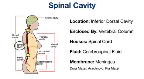
Table of Contents
The Cranial Cavity: A Comprehensive Guide to the Housing of the Brain and Spinal Cord
The human brain, the command center of our entire being, resides within a protective bony structure known as the cranial cavity. This intricate space, part of the larger neurocranium, isn't just a simple container; it's a sophisticated system designed to safeguard this vital organ and facilitate its complex functions. Understanding the cranial cavity, its components, and its relationship with the spinal cord, is crucial to appreciating the delicate balance of the central nervous system.
The Neurocranium: The Bony Fortress
Before delving into the specifics of the cranial cavity itself, let's establish its context within the larger skull. The skull is composed of two main parts: the neurocranium and the viscerocranium. The neurocranium, also known as the braincase, is the protective bony structure that encloses and protects the brain and brainstem. It's formed by eight major bones:
- Frontal Bone: Forms the forehead and part of the anterior cranial fossa.
- Parietal Bones (2): Form the superior and lateral aspects of the cranium.
- Temporal Bones (2): Located on the sides of the skull, housing the inner ear and the structures involved in hearing and balance. They also articulate with the mandible, forming the temporomandibular joint (TMJ).
- Occipital Bone: Forms the posterior part of the skull and contains the foramen magnum, the large opening through which the spinal cord connects to the brainstem.
- Sphenoid Bone: A complex, bat-shaped bone located at the base of the skull. It contributes to several cranial fossae and plays a critical role in the formation of the orbits (eye sockets).
- Ethmoid Bone: A delicate bone located anterior to the sphenoid bone. It contributes to the formation of the nasal cavity and the orbits.
These bones are intricately connected by fibrous joints called sutures, creating a strong, immovable structure. These sutures, while immovable in adults, are flexible in infants, allowing for passage through the birth canal and subsequent brain growth. The sutures are named based on the bones they connect (e.g., the coronal suture connects the frontal and parietal bones).
The Cranial Fossae: Compartmentalizing the Brain
The interior of the neurocranium isn't a single, uniform space. Instead, it's divided into three distinct compartments called cranial fossae: the anterior, middle, and posterior fossae. This compartmentalization helps to protect different parts of the brain and accommodate their varying shapes and sizes.
-
Anterior Cranial Fossa: The most anterior fossa, formed primarily by the frontal bone and parts of the ethmoid and sphenoid bones. It houses the frontal lobes of the brain.
-
Middle Cranial Fossa: A more complex fossa, shaped like a butterfly. It's formed by parts of the sphenoid, temporal, and parietal bones. It houses the temporal lobes and parts of the diencephalon. Key features include the sella turcica, which houses the pituitary gland, and the openings for cranial nerves.
-
Posterior Cranial Fossa: The largest and deepest fossa, formed primarily by the occipital bone and parts of the temporal and parietal bones. It houses the cerebellum, pons, and medulla oblongata (the brainstem). The foramen magnum, the large opening through which the brainstem connects to the spinal cord, is a prominent feature of this fossa.
The Dura Mater: The Brain's Protective Membrane
The brain isn't directly in contact with the bones of the cranial cavity. Instead, it's enveloped by several layers of protective membranes called meninges. The outermost layer is the dura mater, a thick, tough, fibrous membrane. The dura mater has two layers: the periosteal layer (attached to the inner surface of the skull) and the meningeal layer (the deeper layer that forms the actual covering of the brain). Between these layers are important venous sinuses that drain blood from the brain.
The dura mater also forms important structures such as the falx cerebri, a sickle-shaped fold that separates the two cerebral hemispheres, and the tentorium cerebelli, a tent-like structure that separates the cerebrum from the cerebellum. These structures provide additional support and protection for the brain.
The Arachnoid Mater and Pia Mater: Delicate Inner Layers
Beneath the dura mater lie the arachnoid mater and the pia mater. The arachnoid mater is a delicate, web-like membrane that loosely covers the brain. The space between the arachnoid mater and the dura mater is called the subdural space, while the space between the arachnoid mater and the pia mater is the subarachnoid space. This subarachnoid space is filled with cerebrospinal fluid (CSF), a clear, colorless fluid that cushions and protects the brain and spinal cord.
The innermost layer is the pia mater, a thin, transparent membrane that adheres directly to the surface of the brain. It follows the contours of the brain, closely investing its gyri and sulci.
Cerebrospinal Fluid (CSF): The Brain's Protective Fluid
CSF is a critical component of the cranial cavity's protective system. Produced primarily in the choroid plexuses within the ventricles of the brain, CSF circulates through the ventricles, subarachnoid space, and around the spinal cord. Its functions include:
- Cushioning: Absorbing shock and protecting the brain from trauma.
- Buoyancy: Reducing the effective weight of the brain, preventing it from compressing its lower structures.
- Homeostasis: Maintaining a stable chemical environment for the brain.
- Waste Removal: Removing metabolic waste products from the brain.
The Foramina and Cranial Nerves: Communication Pathways
The cranial cavity isn't isolated. Several openings, or foramina, allow for the passage of cranial nerves, blood vessels, and the spinal cord. Twelve pairs of cranial nerves emerge from the brainstem and pass through these foramina to reach their target organs and tissues throughout the head and neck. These nerves control vital functions such as vision, hearing, taste, smell, facial expression, and swallowing.
The Cranial Cavity and the Spinal Cord: A Continuous System
The cranial cavity seamlessly transitions into the vertebral canal, which houses the spinal cord. The foramen magnum marks the transition point, where the brainstem continues as the spinal cord. The spinal cord, like the brain, is protected by meninges and CSF. The meninges continue down the vertebral canal, providing a protective sheath for the spinal cord.
Clinical Significance: Conditions Affecting the Cranial Cavity
Several conditions can affect the cranial cavity, often with serious consequences. These include:
-
Traumatic Brain Injury (TBI): Injury to the brain caused by a blow to the head or a penetrating injury. The severity of TBI can range from mild concussion to severe, life-threatening damage.
-
Intracranial Hemorrhage: Bleeding within the cranial cavity, which can compress brain tissue and cause neurological deficits. Types include epidural hematoma (bleeding between the skull and dura mater), subdural hematoma (bleeding between the dura mater and arachnoid mater), and subarachnoid hemorrhage (bleeding into the subarachnoid space).
-
Meningitis: Infection of the meninges, causing inflammation and potentially severe neurological damage.
-
Brain Tumors: Abnormal growths within the cranial cavity that can compress brain tissue and disrupt normal brain function.
-
Hydrocephalus: A condition characterized by an accumulation of CSF within the cranial cavity, leading to increased intracranial pressure.
Conclusion: A Remarkable Structure
The cranial cavity is far more than just a bony box. It's a sophisticated, intricately designed system that protects the brain, facilitates its functions, and allows for communication with the rest of the body. Understanding its anatomy and physiology is essential for appreciating the delicate balance of the central nervous system and for diagnosing and treating conditions affecting this vital region. The complex interplay of bone, membrane, fluid, and neural pathways within the cranial cavity showcases the remarkable design of the human body and the critical importance of protecting this delicate organ. Further research into the intricacies of the cranial cavity continues to reveal new insights into brain function and protection, contributing to advancements in neurosurgery and neurological care. The ongoing study of the cranial cavity and its components underscores the continuous evolution of our understanding of this essential aspect of human anatomy.
Latest Posts
Latest Posts
-
Investigation Dna Proteins And Sickle Cell Answer Key
Mar 19, 2025
-
Buffer Region On A Titration Curve
Mar 19, 2025
-
Milk Of Magnesia Is Acidic Or Basic
Mar 19, 2025
-
How Does Melting Point Determine Purity
Mar 19, 2025
-
How To Calculate Percent Of Water In A Hydrate
Mar 19, 2025
Related Post
Thank you for visiting our website which covers about Cavity That Contains The Brain And Spinal Cord . We hope the information provided has been useful to you. Feel free to contact us if you have any questions or need further assistance. See you next time and don't miss to bookmark.
