Distinguish The True Pelvis From The False Pelvis
Muz Play
Mar 15, 2025 · 6 min read
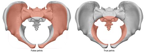
Table of Contents
Distinguishing the True Pelvis from the False Pelvis: A Comprehensive Guide
Understanding the anatomy of the pelvis is crucial for various medical fields, including obstetrics, gynecology, urology, and orthopedics. A key aspect of pelvic anatomy involves differentiating between the true and false pelvis. While both are integral parts of the bony pelvis, they have distinct features, functions, and clinical significances. This comprehensive guide will delve into the anatomical differences, functional distinctions, and clinical implications of distinguishing the true and false pelvis.
Defining the Boundaries: True Pelvis vs. False Pelvis
The human pelvis is a complex bony structure composed of several fused bones: the two hip bones (each comprising the ilium, ischium, and pubis), the sacrum, and the coccyx. This structure is broadly divided into two parts: the false pelvis (also known as the greater pelvis) and the true pelvis (also known as the lesser pelvis). The distinction between these two regions is primarily based on their anatomical boundaries and the role they play in the body.
The False Pelvis: A Supporting Structure
The false pelvis lies superior to the pelvic brim (also known as the linea terminalis or pelvic inlet). It's essentially a part of the abdomen, providing support for the abdominal organs. Its boundaries are less clearly defined than the true pelvis and are formed by:
- Anteriorly: The body of the fifth lumbar vertebra.
- Laterally: The iliac fossae (the broad, shallow depressions on the inner surface of the iliac bones).
- Posteriorly: The lumbar vertebrae (L5 usually being the key landmark).
Key Characteristics of the False Pelvis:
- Shallow and Wide: It's a relatively shallow and broad cavity.
- Limited Obstetrical Significance: It plays a minimal role in childbirth.
- Supports Abdominal Organs: It primarily serves to support the abdominal viscera, including the intestines and the lower parts of the kidneys.
- Muscular Attachment Sites: It provides attachment points for several muscles, including those involved in posture and locomotion.
The True Pelvis: The Birth Canal
The true pelvis, in contrast, lies inferior to the pelvic brim and is the crucial region for childbirth. It's a much more defined, curved, bony canal. Its boundaries are more precise and include:
- Anteriorly: The symphysis pubis.
- Laterally: The ischial bones and the inner surfaces of the hip bones.
- Posteriorly: The sacrum and the coccyx.
Key Characteristics of the True Pelvis:
- Deep and Narrower: Compared to the false pelvis, it is deeper and relatively narrower.
- Obstetrical Significance: This is the passageway through which a fetus travels during childbirth. Its shape and dimensions are crucial in determining the feasibility of vaginal delivery.
- Pelvic Floor Muscles: The pelvic floor muscles form the base of the true pelvis, supporting the pelvic organs and assisting in the process of urination, defecation, and childbirth.
- Pelvic Organs: The true pelvis houses several important organs, including the bladder, rectum, and (in women) the uterus, vagina, and ovaries. The precise relationship of these organs to the bony structure is significant clinically.
Anatomical Differences: A Detailed Comparison
To understand the distinction completely, let's delve deeper into the anatomical differences using comparative tables:
| Feature | False Pelvis | True Pelvis |
|---|---|---|
| Location | Superior to the pelvic brim (linea terminalis) | Inferior to the pelvic brim (linea terminalis) |
| Shape | Shallow and wide | Deep and narrow |
| Boundaries | Iliac fossae, lumbar vertebrae, abdomen | Symphysis pubis, ischial bones, sacrum, coccyx |
| Function | Supports abdominal organs | Forms the birth canal; supports pelvic organs |
| Clinical Relevance | Less significant in obstetrics | Crucial in obstetrics; important in urology, gynecology |
| Clinical Measurements | Not typically measured for obstetrical purposes | Crucial measurements for obstetrical assessment |
Pelvic Inlet vs. Pelvic Outlet: Further Defining the True Pelvis
Within the true pelvis, two crucial boundaries further refine its dimensions and importance: the pelvic inlet and the pelvic outlet.
-
Pelvic Inlet (Superior Pelvic Aperture): This is the superior opening of the true pelvis, formed by the pelvic brim. Its shape and dimensions are critically important in obstetrics. It's defined by the sacral promontory, the arcuate line of the ilium, the pectineal line of the pubic bone, and the superior border of the pubic symphysis.
-
Pelvic Outlet (Inferior Pelvic Aperture): This is the inferior opening of the true pelvis. It is bounded by the coccyx, the ischial tuberosities, and the inferior border of the pubic symphysis. The size and shape of the pelvic outlet are also crucial in determining the feasibility of vaginal delivery.
Clinical Significance: Why the Distinction Matters
The distinction between the true and false pelvis is not merely an anatomical curiosity; it holds significant clinical importance, especially in:
Obstetrics:
- Determining Fetal Passage: The size and shape of the true pelvis are crucial in predicting whether a vaginal delivery is feasible or if a Cesarean section is necessary. Pelvic measurements, such as the diagonal conjugate, interspinous diameter, and intertuberous diameter, are essential in assessing the adequacy of the birth canal.
- Labor and Delivery: The true pelvis acts as the birth canal, and any abnormalities in its shape or dimensions can complicate labor and delivery.
- Postpartum Recovery: Understanding the anatomy of the true pelvis is critical for managing complications such as postpartum hemorrhage or pelvic floor dysfunction.
Gynecology:
- Pelvic Organ Prolapse: The true pelvis supports the pelvic organs. Weakening of the pelvic floor muscles can lead to prolapse of the uterus, bladder, or rectum into the vagina. Understanding the bony structure aids in diagnosing and treating such conditions.
- Gynecological Surgery: Knowledge of pelvic anatomy is essential for various gynecological procedures, ensuring accurate surgical techniques and minimizing complications.
Urology:
- Bladder and Urethral Issues: The true pelvis houses the bladder and urethra. Understanding the relationship between the bony structure and these organs aids in the diagnosis and treatment of conditions such as urinary incontinence and bladder prolapse.
- Prostate Surgery: In men, the prostate gland resides in the true pelvis. Detailed knowledge of pelvic anatomy is critical during prostate surgery.
Orthopedics:
- Pelvic Fractures: Fractures of the pelvic bones, particularly those affecting the true pelvis, can be life-threatening due to potential blood loss and damage to surrounding organs.
- Pelvic Instability: Conditions affecting the sacroiliac joints, which are part of the true pelvis, can lead to pelvic instability and pain.
Imaging Techniques for Pelvic Assessment
Various imaging techniques are used to visualize the pelvis and assess its dimensions and any abnormalities:
- Pelvic X-rays: Provide a clear image of the bony structures of the pelvis, allowing for accurate measurement of the pelvic inlet and outlet.
- Ultrasound: Frequently used in obstetrics to assess fetal position and the size and shape of the maternal pelvis.
- CT scans and MRI: Offer detailed images of the bony structures and soft tissues within the pelvis, providing valuable information in the diagnosis and management of various pelvic conditions.
Conclusion
The distinction between the true and false pelvis is crucial for understanding the anatomy and function of the lower abdomen and pelvis. While the false pelvis primarily supports abdominal organs, the true pelvis serves as the crucial passageway for childbirth and houses vital organs. Accurate knowledge of their anatomical differences and clinical implications is essential for healthcare professionals in various specialties, ultimately leading to improved patient care and outcomes. This detailed understanding allows for appropriate diagnosis, treatment planning, and improved surgical and obstetrical management. Further research continues to refine our understanding of the complex interplay between the bony structures of the pelvis and the various organs it contains, consistently pushing the boundaries of healthcare innovation and advancing patient care.
Latest Posts
Latest Posts
-
High Spin And Low Spin Complexes
Mar 15, 2025
-
Unlike Animal Cells Plant Cells Have
Mar 15, 2025
-
How To Use The Activity Series
Mar 15, 2025
-
Life Cycle Of A Seedless Plant
Mar 15, 2025
-
Proximate And Ultimate Causes Of Behaviour Examples
Mar 15, 2025
Related Post
Thank you for visiting our website which covers about Distinguish The True Pelvis From The False Pelvis . We hope the information provided has been useful to you. Feel free to contact us if you have any questions or need further assistance. See you next time and don't miss to bookmark.
