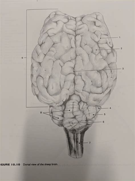
Dorsal View Of The Sheep Brain
Muz Play
Apr 02, 2025 · 6 min read

Table of Contents
A Comprehensive Exploration of the Sheep Brain's Dorsal View
The sheep brain, a readily available and ethically sourced model for studying mammalian neuroanatomy, offers a fascinating glimpse into the intricate structures of the central nervous system. While dissection allows for a complete exploration, focusing on a specific view, like the dorsal view, provides a focused understanding of key anatomical features. This article offers a detailed examination of the sheep brain's dorsal surface, covering its major structures, their functions, and clinical significance.
Understanding the Dorsal View: A Starting Point
The dorsal view, also known as the superior view, presents a "top-down" perspective of the brain. It provides a crucial overview of the cerebrum's major lobes, the cerebellum, and the connecting structures. Understanding this view is fundamental to grasping the overall organization and functionality of the sheep brain, and by extension, the mammalian brain as a whole. It’s essential to note that the sheep brain exhibits a high degree of similarity to the human brain, making it an excellent comparative model.
Key Structures Visible in the Dorsal View
Several prominent structures are clearly visible from the dorsal perspective:
-
Cerebrum: This is the largest part of the sheep brain, responsible for higher-level cognitive functions, including learning, memory, and voluntary movement. Its highly convoluted surface, characterized by gyri (ridges) and sulci (grooves), significantly increases surface area, accommodating the vast number of neurons. On the dorsal view, the cerebrum dominates, showcasing its two cerebral hemispheres separated by the longitudinal fissure.
-
Cerebral Hemispheres: The two symmetrical halves of the cerebrum are distinctly visible. The corpus callosum, a large bundle of nerve fibers connecting the two hemispheres and enabling interhemispheric communication, is partially visible in a midsagittal section (although not fully visible from a purely dorsal view).
-
Frontal Lobe: Located at the anterior (front) portion of the cerebrum, the frontal lobe plays a critical role in executive functions, planning, decision-making, and voluntary motor control. Its size and position are clearly evident from the dorsal aspect.
-
Parietal Lobe: Situated posterior to the frontal lobe, the parietal lobe is involved in processing sensory information, particularly touch, temperature, pain, and spatial awareness. Its boundaries are readily identifiable from the dorsal view, although some portions might be obscured by overlying structures.
-
Temporal Lobe: Partially visible from the dorsal view (more prominently seen from lateral views), the temporal lobe is crucial for auditory processing, memory consolidation, and language comprehension. The superior temporal gyrus, a major component, might be partially visible depending on the preparation of the brain.
-
Occipital Lobe: Located at the posterior (rear) end of the cerebrum, the occipital lobe is primarily dedicated to visual processing. While its full extent isn't fully visible from a strictly dorsal view, its posterior location is readily apparent.
-
Cerebellum: Situated beneath the occipital lobe and posterior to the brainstem, the cerebellum is responsible for coordinating movement, balance, and posture. Its dorsal surface shows distinct folia (leaf-like folds), creating a textured appearance. The vermis, a midline structure of the cerebellum, is clearly visible as a central ridge separating the cerebellar hemispheres.
Deeper Dive into Specific Structures and their Functions
Let's delve deeper into the functional roles of the key structures observable from the dorsal view.
The Cerebrum: A Command Center
The cerebrum’s intricate structure reflects its complex functions. Its gyri and sulci maximize the surface area, enabling the dense packing of neurons and facilitating advanced cognitive capabilities. The dorsal view allows a clear visualization of the relative sizes of the lobes, suggesting the weighting of different cognitive functions in sheep. The prominent frontal lobe, for instance, indicates a likely sophisticated level of motor control and planning behaviors.
Frontal Lobe Functions in Sheep:
While we can't directly infer human-like cognitive processes, the frontal lobe's size in sheep suggests crucial roles in:
- Spatial navigation: Sheep rely heavily on spatial memory for foraging and social interactions. The frontal lobe is heavily involved in processing spatial information.
- Predator avoidance: Quick decision-making and escape strategies are vital for survival. The frontal lobe facilitates these responses.
- Social hierarchy: Sheep live in complex social structures, requiring sophisticated social cognition. The frontal lobe contributes to understanding and navigating social hierarchies.
The Cerebellum: Master of Motor Coordination
The cerebellum's dorsal view showcases its folia, which dramatically increase the surface area for neuronal connections responsible for fine-tuning motor control. Observing the cerebellum's size and relative proportion compared to other brain structures provides insights into the sheep's locomotor abilities and coordination skills.
Cerebellar Functions in Sheep:
- Precise locomotion: Sheep demonstrate considerable agility and balance. The cerebellum is instrumental in coordinating their movements.
- Maintaining posture: The cerebellum's role in maintaining balance and posture is crucial, particularly on uneven terrain.
- Rapid adjustments: Sheep need to quickly adjust their gait and posture in response to changes in their environment. The cerebellum facilitates these rapid adjustments.
The Connections: Integrating Information
Although not fully visible from a purely dorsal perspective, the connections between different brain regions are crucial for information integration. The corpus callosum, visible in a slightly shifted view or section, highlights the interhemispheric communication essential for coordinating activities across the brain.
Clinical Significance: Linking Structure to Function
Studying the dorsal view of the sheep brain provides valuable insights into neurological disorders and their impact. Any anomalies or deviations from the typical morphology can suggest underlying pathologies. Comparative studies between healthy and diseased brains highlight the correlation between structure and function, aiding diagnosis and understanding of neurological conditions.
Applications in Veterinary Medicine
The sheep brain serves as a relevant model for understanding neurological diseases in other ruminants. Observing deviations in the dorsal view, such as abnormal gyri formation, asymmetries in the hemispheres, or size discrepancies in the cerebellum, could indicate developmental abnormalities or neurological conditions.
Implications for Human Neuroscience
The similarities between sheep and human brain structures make the sheep brain a valuable model for exploring human neuroanatomy and neurophysiology. Research on the sheep brain can enhance our understanding of human brain development, cognitive processes, and neurological disorders. Studying the comparative morphology of different brain regions aids in developing potential therapeutic strategies.
Practical Tips for Observing the Dorsal View
When examining a sheep brain's dorsal view, consider the following:
- Brain Preparation: Ensure the brain is properly fixed and preserved to maintain its integrity and anatomical details.
- Appropriate Lighting: Good lighting is essential for clear visualization of the brain's surface features.
- Magnification: Using a dissecting microscope or magnifying glass can reveal finer details of the gyri and sulci.
- Comparative Analysis: Compare your observations with anatomical atlases and diagrams to ensure accurate identification of structures.
Conclusion: A Window into the Mammalian Brain
The dorsal view of the sheep brain, while only one perspective, provides a fundamental framework for understanding the intricate architecture and functional organization of the mammalian brain. By carefully observing and analyzing this view, we can gain valuable insights into the complexities of neurological systems, with implications for both veterinary and human neuroscience. Further exploration of other perspectives, coupled with histological and functional studies, provides a more comprehensive understanding of this remarkable organ. The sheep brain, therefore, remains a powerful tool for advancing our knowledge of brain function and pathology.
Latest Posts
Latest Posts
-
Chloroplast In Plant Cell Under Microscope
Apr 03, 2025
-
Contribution Of John Newlands In Periodic Table
Apr 03, 2025
-
Measure Of The Amount Of Matter
Apr 03, 2025
-
Bases Produce Which Ions In Aqueous Solution
Apr 03, 2025
-
An Atom That Has Lost Or Gained An Electron
Apr 03, 2025
Related Post
Thank you for visiting our website which covers about Dorsal View Of The Sheep Brain . We hope the information provided has been useful to you. Feel free to contact us if you have any questions or need further assistance. See you next time and don't miss to bookmark.
