How Does A Tendon Sheath Differ From A Bursa
Muz Play
Mar 24, 2025 · 5 min read
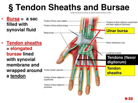
Table of Contents
How Does a Tendon Sheath Differ From a Bursa?
Both tendon sheaths and bursae are crucial components of the musculoskeletal system, acting as cushions and reducing friction during movement. While they share the common goal of facilitating smooth joint motion, their anatomical structures, locations, and functions differ significantly. Understanding these distinctions is key to comprehending musculoskeletal injuries and their appropriate treatments.
Understanding Tendon Sheaths
Tendon sheaths are specialized structures that envelop tendons, primarily those subjected to high levels of friction and repetitive movement. They are essentially elongated, tube-like bursae that completely enclose the tendon throughout its course. This complete encasement is a key distinguishing feature from bursae.
Anatomy of a Tendon Sheath
A tendon sheath comprises two layers:
-
Outer Fibrous Layer (Vagina Fibrosa): This layer is composed of dense, fibrous connective tissue. It provides structural support and protection to the tendon within. It’s relatively strong and less pliable than the inner layer.
-
Inner Synovial Layer (Vagina Synovialis): This layer is a delicate membrane that lines the inner surface of the fibrous layer. It produces synovial fluid, a viscous lubricant that minimizes friction between the tendon and the sheath. This fluid is essential for smooth tendon gliding. The synovial layer is divided into two further components: a visceral layer which directly adheres to the tendon and a parietal layer which lines the inner surface of the fibrous sheath. Between these two layers is a potential space that is filled with synovial fluid.
Location and Function of Tendon Sheaths
Tendon sheaths are primarily found in areas where tendons undergo significant bending or are subjected to considerable forces. Common locations include:
- Wrist: The tendons of the flexor and extensor muscles of the hand are frequently enveloped by tendon sheaths.
- Fingers: Tendons in the fingers, particularly those involved in flexion and extension, are also often surrounded by sheaths.
- Ankle: Tendons around the ankle, crucial for foot movement, can have tendon sheaths.
- Toes: Similar to the fingers, tendons in the toes can also be enclosed by these structures.
The primary function of a tendon sheath is to reduce friction during tendon movement. The synovial fluid acts as a highly effective lubricant, allowing tendons to glide smoothly within the sheath, preventing wear and tear. This is particularly crucial in areas of repetitive movement.
Understanding Bursae
Bursae are small, fluid-filled sacs located strategically around joints and between tendons and bones. They act as cushions, reducing friction between moving structures. Unlike tendon sheaths, they don't completely encase a tendon.
Anatomy of a Bursa
A bursa is essentially a closed sac lined by a synovial membrane. This membrane produces synovial fluid, which lubricates the bursa and minimizes friction. The fluid within the bursa is similar in composition to the synovial fluid found in joints. The exterior of the bursa may be reinforced by fibrous connective tissue.
Location and Function of Bursae
Bursae are distributed throughout the body, often located in areas prone to friction:
-
Shoulders: Several bursae are found in the shoulder joint, protecting the tendons of the rotator cuff muscles. The subacromial bursa, for example, lies between the acromion process of the scapula and the supraspinatus tendon.
-
Elbows: Bursae cushion the movement of tendons around the elbow joint.
-
Hips: Bursae are found in the hip region to facilitate movement and reduce friction. The trochanteric bursa is a notable example.
-
Knees: The prepatellar bursa lies in front of the kneecap and helps to reduce friction between the patella and the skin.
-
Feet: Bursae provide cushioning in the feet, especially in areas that bear weight.
The primary function of a bursa is to reduce friction between different structures that move against each other. This friction reduction prevents inflammation and damage to the underlying tissues.
Key Differences Between Tendon Sheaths and Bursae
The following table summarizes the key distinctions between tendon sheaths and bursae:
| Feature | Tendon Sheath | Bursa |
|---|---|---|
| Structure | Elongated, tube-like; two layers (fibrous & synovial) | Closed sac; lined by synovial membrane |
| Location | Completely encloses tendons | Located between tendons, bones, and skin |
| Function | Reduces friction between tendon and surrounding structures | Reduces friction between adjacent structures |
| Fluid | Synovial fluid within the double-layered sheath | Synovial fluid within the bursa itself |
| Tendon Relation | Completely surrounds tendon | Does not completely encase the tendon |
| Shape | Tubular or cylindrical | Typically flattened or sac-like |
Clinical Significance: Inflammation and Disorders
Both tendon sheaths and bursae are susceptible to inflammation, leading to painful and debilitating conditions.
Tendon Sheath Disorders
-
Tenosynovitis: Inflammation of the tendon sheath, often caused by overuse, trauma, or infection. Symptoms include pain, swelling, and crepitus (a crackling sound during movement). De Quervain's tenosynovitis is a common example affecting the thumb tendons.
-
Stenosing Tenosynovitis (Trigger Finger): A condition where the tendon sheath becomes thickened and inflamed, causing the tendon to catch and snap during movement.
Bursa Disorders
-
Bursitis: Inflammation of a bursa, usually due to overuse, trauma, or infection. Symptoms include pain, swelling, and tenderness over the affected bursa. Prepatellar bursitis (housemaid's knee) is a common example.
-
Septic Bursitis: A serious condition where a bursa becomes infected, often requiring medical intervention.
Treatment Approaches
Treatment for tendon sheath and bursa disorders typically focuses on rest, ice, compression, and elevation (RICE protocol). Other treatments may include:
- Non-steroidal anti-inflammatory drugs (NSAIDs): To reduce pain and inflammation.
- Corticosteroid injections: To reduce inflammation directly in the affected area.
- Physical therapy: To improve range of motion and strengthen surrounding muscles.
- Surgery: In severe cases, surgery may be necessary to repair damaged tissues or release constricted tendons.
Conclusion
While both tendon sheaths and bursae share the vital role of reducing friction in the musculoskeletal system, their anatomical structures and locations distinguish them. Understanding these differences is essential for accurate diagnosis and effective management of conditions affecting these crucial structures. The complete encasement of a tendon by a sheath versus the strategic placement of a bursa highlights their distinct contributions to efficient and pain-free movement. Recognizing these differences is paramount for healthcare professionals in diagnosing and treating various musculoskeletal injuries and ailments. Further research continues to refine our understanding of the intricate workings of these crucial anatomical components and their roles in maintaining musculoskeletal health.
Latest Posts
Latest Posts
-
Do Fingernails And Hair Have Chitin In Them
Mar 26, 2025
-
Separation Of Variables Partial Differential Equations Examples
Mar 26, 2025
-
What Are The 3 Properties Of Water
Mar 26, 2025
-
Learn The Parts Of The Animal Cell Coloring Answer Key
Mar 26, 2025
-
Why Is Nucleus Called The Control Center Of The Cell
Mar 26, 2025
Related Post
Thank you for visiting our website which covers about How Does A Tendon Sheath Differ From A Bursa . We hope the information provided has been useful to you. Feel free to contact us if you have any questions or need further assistance. See you next time and don't miss to bookmark.
