Learn The Parts Of The Animal Cell Coloring Answer Key
Muz Play
Mar 26, 2025 · 5 min read
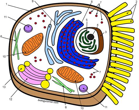
Table of Contents
Learn the Parts of the Animal Cell: A Comprehensive Coloring Guide and Answer Key
Learning about animal cells can be engaging and fun, especially when combined with hands-on activities like coloring. This comprehensive guide will not only take you through the key components of an animal cell but also provide you with a detailed answer key to a fun coloring exercise. Let's dive in!
Understanding the Animal Cell: A Microscopic World
Animal cells are the fundamental building blocks of animals, including humans. Unlike plant cells, they lack a rigid cell wall and chloroplasts. However, they possess a variety of other organelles that work together to maintain the cell's structure and function. Mastering the parts of an animal cell is crucial to understanding biology and related fields.
Key Components of the Animal Cell:
1. Cell Membrane (Plasma Membrane): This is the outer boundary of the cell, a selectively permeable barrier that regulates what enters and exits. Think of it as a gatekeeper controlling the flow of substances. Function: Protects the cell, regulates transport of materials.
2. Cytoplasm: This is the jelly-like substance that fills the cell. It's the site where many cellular processes occur. Function: Supports and suspends organelles, site of many metabolic reactions.
3. Nucleus: Often called the "control center," the nucleus houses the cell's genetic material (DNA). It's enclosed by a double membrane called the nuclear envelope. Function: Contains DNA, controls gene expression, regulates cell activities.
4. Nucleolus: Found within the nucleus, the nucleolus is involved in ribosome production. Function: Ribosome synthesis.
5. Ribosomes: These are tiny structures responsible for protein synthesis. They can be free-floating in the cytoplasm or attached to the endoplasmic reticulum. Function: Protein synthesis.
6. Endoplasmic Reticulum (ER): A network of membranes extending throughout the cytoplasm. There are two types:
* **Rough Endoplasmic Reticulum (RER):** Studded with ribosomes, it's involved in protein synthesis and modification. **Function:** Protein synthesis, modification, and transport.
* **Smooth Endoplasmic Reticulum (SER):** Lacks ribosomes and is involved in lipid synthesis and detoxification. **Function:** Lipid synthesis, detoxification, calcium storage.
7. Golgi Apparatus (Golgi Body): This organelle processes and packages proteins and lipids for transport within or outside the cell. Think of it as the cell's post office. Function: Processes, packages, and distributes proteins and lipids.
8. Mitochondria: Often called the "powerhouses" of the cell, mitochondria generate energy (ATP) through cellular respiration. Function: ATP production through cellular respiration.
9. Lysosomes: These are membrane-bound sacs containing digestive enzymes that break down waste materials and cellular debris. Function: Waste breakdown, cellular recycling.
10. Vacuoles: Storage sacs for water, nutrients, and waste products. Animal cells typically have smaller vacuoles compared to plant cells. Function: Storage of water, nutrients, and waste.
11. Centrosomes: These are regions near the nucleus that organize microtubules, which are important for cell division. Function: Microtubule organization, cell division.
Animal Cell Coloring Activity: A Step-by-Step Guide
Now, let's put our knowledge to the test with a fun coloring activity! Below is a simplified representation of an animal cell; use the descriptions above to color and label the different parts.
(Insert a simple line drawing of an animal cell here. The drawing should be clear and large enough to color, including clearly visible representations of the nucleus, nucleolus, cell membrane, cytoplasm, ribosomes (both free and on RER), RER, SER, Golgi apparatus, mitochondria, lysosomes, vacuoles, and centrosomes.)
Coloring Suggestions:
- Cell Membrane: Light Blue
- Cytoplasm: Light Yellow
- Nucleus: Dark Pink
- Nucleolus: Darker Pink
- Ribosomes: Purple
- Rough Endoplasmic Reticulum (RER): Darker Blue
- Smooth Endoplasmic Reticulum (SER): Light Green
- Golgi Apparatus: Orange
- Mitochondria: Red
- Lysosomes: Dark Green
- Vacuoles: Light Purple
- Centrosomes: Brown
Remember to label each part clearly with its name!
Animal Cell Coloring Answer Key
This section provides a detailed answer key for the coloring activity. Remember, the colors are suggestions, and your creativity is encouraged! The key focuses on the accurate identification and labeling of each organelle.
(Replicate the line drawing of the animal cell here, but this time, each organelle is clearly labeled with its name. The colors used should correspond to the suggestions provided earlier, or a similar scheme to maintain visual clarity and consistency.)
Detailed Labels:
- Cell Membrane: The outer boundary of the cell, a thin, selectively permeable membrane.
- Cytoplasm: The jelly-like substance filling the cell, containing various organelles.
- Nucleus: The control center of the cell, containing the genetic material (DNA).
- Nucleolus: A dark-staining region within the nucleus, involved in ribosome production.
- Ribosomes (Free and on RER): Small, granular structures responsible for protein synthesis. Note the difference in location: free-floating or attached to the RER.
- Rough Endoplasmic Reticulum (RER): A network of membranes studded with ribosomes, involved in protein synthesis and modification.
- Smooth Endoplasmic Reticulum (SER): A network of membranes without ribosomes, involved in lipid synthesis and detoxification.
- Golgi Apparatus: A stack of flattened sacs that processes and packages proteins and lipids for secretion.
- Mitochondria: The powerhouses of the cell, generating energy (ATP) through cellular respiration.
- Lysosomes: Membrane-bound sacs containing digestive enzymes for waste breakdown.
- Vacuoles: Storage sacs for water, nutrients, and waste products.
- Centrosomes: Regions near the nucleus that organize microtubules essential for cell division.
Beyond the Coloring Page: Further Exploration
Completing the coloring activity is just the beginning of your journey into the fascinating world of animal cells. Here are some avenues for further exploration:
- Microscopy: If possible, observe prepared slides of animal cells under a microscope. This provides a real-world perspective of the structures you've colored.
- Interactive Simulations: Numerous online resources offer interactive simulations of animal cells, allowing you to explore their structure and function in a dynamic environment.
- Research Projects: Dive deeper into specific organelles. Research their functions in more detail, exploring related diseases or advancements in research.
- Comparative Cell Biology: Compare and contrast animal cells with plant cells, highlighting the similarities and differences.
Conclusion
Understanding the structure and function of animal cells is a cornerstone of biological knowledge. By combining a hands-on approach like coloring with detailed explanations and further exploration, you can significantly enhance your understanding and appreciation of these remarkable microscopic entities. The coloring activity and answer key provided here serve as valuable tools to make learning fun and effective. Remember to always be curious and keep exploring the wonders of the microscopic world!
Latest Posts
Latest Posts
-
Electron Transport Chain Final Electron Acceptor
Mar 28, 2025
-
Select The Components Of A Fatty Acid
Mar 28, 2025
-
Is Salt A Pure Substance Or A Mixture
Mar 28, 2025
-
What Is The Subscript In Chemistry
Mar 28, 2025
-
What Is The Ph Of Salt
Mar 28, 2025
Related Post
Thank you for visiting our website which covers about Learn The Parts Of The Animal Cell Coloring Answer Key . We hope the information provided has been useful to you. Feel free to contact us if you have any questions or need further assistance. See you next time and don't miss to bookmark.
