How To Create A Wet Mount Slide
Muz Play
Mar 21, 2025 · 6 min read
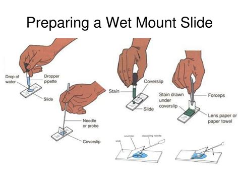
Table of Contents
How to Create a Wet Mount Slide: A Comprehensive Guide
Creating a wet mount slide is a fundamental technique in microscopy, allowing for the observation of living organisms and specimens in their natural aqueous environment. This comprehensive guide will walk you through the process, covering everything from choosing the right materials to mastering the technique for optimal viewing. We'll also delve into troubleshooting common issues and explore advanced techniques to enhance your microscopic observations.
Understanding Wet Mounts: Why and When to Use Them
A wet mount slide involves suspending a specimen in a drop of liquid on a microscope slide, then covering it with a coverslip. This simple preparation offers several advantages:
-
Observing Living Organisms: Wet mounts are ideal for studying living microorganisms like bacteria, protists, and algae in their natural state. The aqueous environment allows for observation of movement, feeding, and other dynamic processes.
-
Maintaining Specimen Integrity: Unlike other preparation methods that may require staining or fixing, wet mounts minimize disruption to the specimen's structure and cellular components. This is particularly crucial for delicate specimens.
-
Simplicity and Speed: Wet mount preparation is quick and easy, requiring minimal equipment and expertise. This makes it an accessible technique for beginners and experienced microscopists alike.
-
Cost-Effectiveness: The materials required for wet mount preparation are inexpensive and readily available, making it a budget-friendly technique.
Materials You Will Need
Before you begin, gather the necessary materials. Having everything prepared in advance ensures a smooth and efficient process:
-
Microscope Slides: Choose clean, grease-free slides. Pre-cleaned slides are readily available commercially.
-
Coverslips: These are thin, square or rectangular pieces of glass designed to cover the specimen. Sizes typically range from 18mm x 18mm to 24mm x 24mm. Ensure they are clean and free of dust or debris.
-
Specimen: This could be anything from a drop of pond water containing microorganisms to a prepared sample of cells or tissues.
-
Dropper or Pipette: Used for carefully dispensing the mounting medium.
-
Mounting Medium: The choice of mounting medium depends on the specimen and the desired observation time. Common options include:
- Water: Simple, readily available, and suitable for short-term observations.
- Saline Solution (0.9% NaCl): Isotonic solution mimicking the body's salinity, beneficial for observing living cells.
- Culture Medium: If observing microorganisms from a culture, using the original culture medium helps maintain the specimen's viability.
- Glycerin: Provides a longer-lasting mount, preventing drying and allowing for more extended observations.
-
Dissecting Needle or Forceps (Optional): Useful for manipulating the specimen onto the slide.
-
Lens Paper or Kimwipes: Used for cleaning slides and coverslips.
-
Microscope: Naturally, you'll need a microscope to view your wet mount!
Step-by-Step Guide to Creating a Wet Mount Slide
Follow these steps for creating a high-quality wet mount slide:
-
Prepare Your Slide: Ensure your microscope slide is clean and free of fingerprints or dust. Gently wipe it with lens paper if necessary.
-
Add the Mounting Medium: Using a dropper or pipette, place a small drop (approximately the size of a grain of rice) of your chosen mounting medium in the center of the slide.
-
Introduce the Specimen: Carefully introduce your specimen into the drop of mounting medium. If using a dissecting needle or forceps, handle the specimen gently to avoid damaging it. If working with a liquid sample like pond water, a single drop will suffice. For solid samples, you may need to carefully tease apart a small portion.
-
Lower the Coverslip: Hold the coverslip at a 45-degree angle, with one edge touching the slide near the edge of the mounting medium drop. Slowly lower the coverslip onto the slide, allowing the medium to spread evenly under the coverslip. Avoid trapping air bubbles. If bubbles are present, gently tap the coverslip with the end of a pencil or pen to dislodge them.
-
Remove Excess Liquid: If there's excess liquid that has overflowed, carefully blot it away using a Kimwipe. This prevents the liquid from interfering with the microscope's objective lens.
-
Examine Under the Microscope: Carefully place the slide onto the microscope stage and secure it with the stage clips. Start with a low-power objective lens to locate your specimen, then switch to higher magnifications for detailed observation.
Troubleshooting Common Wet Mount Issues
Even with careful preparation, you might encounter some challenges. Here are solutions to common problems:
-
Air Bubbles: As mentioned earlier, gently tapping the coverslip can dislodge air bubbles. Alternatively, you can try using less mounting medium and carefully lowering the coverslip.
-
Coverslip Drift: If the coverslip moves or slides during observation, ensure it's properly secured with a small amount of mounting medium. Using a thin layer of petroleum jelly around the edges of the coverslip can provide additional adhesion but use sparingly to prevent contaminating the specimen.
-
Specimen Too Thick or Dense: If the specimen is too thick, it can make focusing difficult. Try using a smaller amount of specimen or using a more diluted sample.
-
Specimen Drying Out: For extended observations, use a longer-lasting mounting medium like glycerin or seal the edges of the coverslip with clear nail polish (applied carefully to the edges only).
-
Contamination: Always use clean slides and coverslips. Sterilize your equipment if working with potentially hazardous samples.
Advanced Wet Mount Techniques
For more advanced applications, consider these techniques:
-
Staining: Adding a stain can enhance contrast and visibility, particularly for transparent specimens. However, note that this will usually kill any living specimens. Common stains include methylene blue, iodine, and crystal violet. Apply the stain to one edge of the coverslip, allowing it to diffuse under the coverslip through capillary action.
-
Hanging Drop Mount: For specimens that are very motile or sensitive to pressure, a hanging drop mount is preferred. This involves suspending the drop of medium and the specimen from the underside of the coverslip. This provides a more natural environment for the specimen and minimizes distortion.
-
Ringed Mounts: For long-term preservation, the edges of the coverslip can be sealed with a sealant such as petroleum jelly or specialized mounting media. This will prevent the evaporation of the mounting liquid and help preserve your specimen for a longer duration.
-
Micrometry: To obtain quantitative data, you can incorporate a micrometer slide into your observations. This allows for precise measurements of the specimen's size and other features.
Conclusion: Mastering the Art of the Wet Mount
Creating a wet mount slide is a foundational skill in microscopy. By following this comprehensive guide and mastering the techniques, you can unlock a world of microscopic wonders and observe the intricacies of living organisms and specimens. Remember that practice is key – the more you create wet mounts, the more proficient you will become. Don't be afraid to experiment with different mounting media, specimens, and techniques to optimize your observations and expand your microscopic explorations. Happy observing!
Latest Posts
Latest Posts
-
What Happens During The Reduction Stage Of The Calvin Cycle
Mar 27, 2025
-
Is Solid To Liquid Endothermic Or Exothermic
Mar 27, 2025
-
What Does A Negative Enthalpy Mean
Mar 27, 2025
-
Divides The Body Into Anterior And Posterior Portions
Mar 27, 2025
-
Claim Of Fact Value And Policy
Mar 27, 2025
Related Post
Thank you for visiting our website which covers about How To Create A Wet Mount Slide . We hope the information provided has been useful to you. Feel free to contact us if you have any questions or need further assistance. See you next time and don't miss to bookmark.
