How To Make A Wet Mount
Muz Play
Apr 05, 2025 · 7 min read

Table of Contents
How to Make a Wet Mount: A Comprehensive Guide for Beginners and Experts
Creating a wet mount is a fundamental technique in microscopy, offering a simple yet effective way to observe living organisms and specimens in their natural, aqueous environment. Whether you're a student conducting a biology experiment, a hobbyist exploring the microscopic world, or a professional researcher needing a quick sample preparation method, mastering the wet mount technique is crucial. This comprehensive guide covers everything you need to know, from choosing the right equipment to troubleshooting common issues, ensuring you create high-quality, clear wet mounts every time.
Understanding Wet Mounts: The Basics
A wet mount, also known as a temporary mount, involves suspending a specimen in a drop of liquid (usually water or a saline solution) on a microscope slide, then covering it with a coverslip. This simple preparation allows for the observation of living organisms in their natural state, minimizing the artifacts often introduced by more complex preparation techniques. The liquid provides a medium for the specimen, preventing it from drying out and preserving its natural shape and movement.
The key advantages of using a wet mount include:
- Simplicity: Wet mounts are incredibly easy to prepare, requiring minimal equipment and expertise.
- Observation of Living Organisms: This technique is ideal for observing the movement and behavior of live specimens like microorganisms, protozoa, and algae.
- Speed: Preparation time is minimal, making it perfect for quick observations.
- Cost-Effectiveness: Wet mounts require minimal materials, keeping costs low.
Essential Equipment and Materials
Before you begin, gather the necessary materials. Having everything prepared beforehand streamlines the process and prevents interruptions. You'll need:
- Microscope Slides: Standard microscope slides are rectangular glass slides, usually 75mm x 25mm. Clean slides are essential for optimal viewing.
- Coverslips: These are thin, square pieces of glass, typically ranging from 18mm x 18mm to 22mm x 22mm. The size should be appropriate for the slide and the specimen. Avoid using coverslips that are too large or too small.
- Pipettes or Droppers: These are used to transfer small quantities of liquid, ensuring precise application of the mounting medium. Pasteur pipettes are excellent for this purpose.
- Mounting Medium: Distilled water is the most common mounting medium, providing a neutral environment for many specimens. Other options include saline solutions (for maintaining osmotic balance), physiological saline (0.9% NaCl), or specialized stains and mounting media depending on the sample and the desired observation.
- Specimen: This could be anything from pond water teeming with microorganisms to a prepared sample of plant cells or a single insect leg.
- Forceps or Tweezers: Useful for handling small specimens or delicate materials.
- Dissecting Needle or Probe: Helpful for manipulating specimens on the slide.
- Lens Paper or Kimwipes: Essential for cleaning the microscope slides and coverslips before and after use.
- Microscope: Naturally, a microscope is the centerpiece of this process!
Step-by-Step Guide to Creating a Perfect Wet Mount
Now, let's delve into the process of creating a high-quality wet mount:
1. Prepare Your Workspace: Ensure you have a clean and well-lit area to minimize the risk of contamination and enhance visibility.
2. Clean Your Slides and Coverslips: Using lens paper or Kimwipes, gently clean the microscope slides and coverslips. Dust and fingerprints can significantly interfere with your observations.
3. Prepare Your Specimen: Depending on the specimen, this may involve collecting a sample from its natural environment (like pond water), preparing a tissue sample, or using a prepared slide. If necessary, use forceps or a dissecting needle to carefully place the specimen onto the center of the slide.
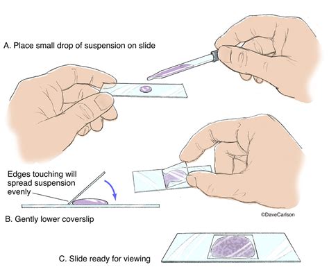
4. Apply the Mounting Medium: Using a pipette or dropper, add a single, small drop of your chosen mounting medium (usually distilled water) directly onto the center of the slide, where the specimen is located. Avoid adding too much liquid, as this can overflow under the coverslip and make observation difficult. The amount should ideally be only enough to suspend the specimen adequately.
5. Carefully Lower the Coverslip: This is a crucial step. Hold the coverslip at a 45-degree angle, with one edge touching the edge of the drop of mounting medium. Slowly lower the coverslip onto the slide, allowing the liquid to spread evenly underneath. Avoid trapping air bubbles, which can significantly obstruct your view.
6. Remove Excess Liquid: If there's an excessive amount of liquid that has spread beyond the edges of the coverslip, gently blot the excess liquid away with a piece of absorbent paper or a Kimwipe. Be careful not to move or disrupt the coverslip.
7. Observe Under the Microscope: Carefully place your wet mount onto the stage of your microscope and begin observation at low magnification. Gradually increase the magnification as needed to achieve the desired level of detail.
Troubleshooting Common Wet Mount Problems
Even with careful preparation, certain issues can arise when creating wet mounts. Let's examine some common problems and their solutions:
-
Air Bubbles: Air bubbles are a common nuisance that can obscure your view. To minimize their formation, carefully lower the coverslip at an angle, ensuring it makes even contact with the slide. Gentle tapping on the coverslip can sometimes dislodge small bubbles. If excessive bubbles remain, remake the wet mount.
-
Specimen Too Dry: If your specimen appears dry or shrunken, you may not have added enough mounting medium. If this occurs during observation, carefully add a small drop of additional liquid at the edge of the coverslip using a pipette, allow a minute or so for it to permeate, and resume your observations.
-
Specimen Too Wet: If there’s excessive liquid, gently wick away some of the excess with absorbent paper. Again, take care not to move the coverslip.
-
Coverslip Movement: If the coverslip keeps moving, this can hinder your observation. Make sure the coverslip adheres securely to the slide, and consider using a slightly smaller coverslip to reduce movement. You can also very lightly apply clear nail polish to the edges of the coverslip (allowing the polish to dry thoroughly before using the slide) to secure it in place – although this is only suitable for observations that do not require long-term storage.
-
Unclear Image: This can be due to several factors, including dirty slides or coverslips, excessive air bubbles, a poor-quality specimen, or incorrect microscope settings. Check and correct the above issues before resuming your observations.
Advanced Techniques and Considerations
While the basic wet mount technique is straightforward, several advanced techniques can enhance your observations:
-
Staining: Adding stains to your mounting medium can enhance the visibility of specific structures within the specimen. For example, methylene blue is commonly used to stain cells, making their nuclei and other structures more visible. However, stains may kill the organisms and will alter their natural state, which should be taken into account.
-
Using Different Mounting Media: While water is a common choice, other media such as saline solutions or specialized mounting fluids are ideal for specific specimens or observations.
-
Creating Hanging Drop Mounts: This variation is particularly useful for observing the motility of microorganisms. A small drop of the specimen suspension is placed on an inverted coverslip, which is then carefully placed onto a well slide or depression slide to minimize evaporation and enable a longer observation duration.
-
Using a Phase-Contrast Microscope: For specimens that have very little contrast, a phase-contrast microscope can dramatically improve the visibility of details.
-
Microscope Calibration and Maintenance: Regular microscope maintenance and accurate calibration of your microscope will ensure your observations are clear and reliable.
Applications of Wet Mounts
The applications of wet mounts extend far beyond simple classroom experiments:
-
Biology Education: Wet mounts are a staple in biology education, allowing students to observe living cells, microorganisms, and other biological samples.
-
Water Quality Analysis: Observing microorganisms in water samples can provide valuable insights into water quality and potential contamination.
-
Parasitology: Wet mounts are used in the identification of parasites in clinical samples.
-
Medical Diagnosis: While not primary methods, they can provide a quick assessment for certain pathogens under specific conditions.
-
Environmental Monitoring: Studying the diversity of microorganisms in various environments helps assess environmental health.
-
Hobbyist Microscopy: Many enthusiasts use wet mounts to explore the wonders of the microscopic world, such as examining pollen, diatoms, or the intriguing world found within a drop of pond water.
Conclusion
The wet mount technique is a cornerstone of microscopy. While seemingly simple, mastering this fundamental technique allows you to unlock the hidden world of microscopy. By following these steps, troubleshooting common issues, and exploring advanced techniques, you'll be well-equipped to create high-quality wet mounts for diverse applications, whether for educational purposes, scientific research, or personal exploration of the fascinating microscopic realm. Remember that practice is key. With enough experimentation, creating flawless wet mounts will become second nature, ensuring your microscope journey is both rewarding and insightful.
Latest Posts
Latest Posts
-
Are Acid Fast Bacteria Gram Negative Or Positive
Apr 05, 2025
-
How To Make A Titration Curve In Excel
Apr 05, 2025
-
Which Describes The Complex Carbohydrate Cellulose
Apr 05, 2025
-
After Glycolysis The Pyruvate Molecules Go To The
Apr 05, 2025
-
Converting Polar Equations To Cartesian Equations
Apr 05, 2025
Related Post
Thank you for visiting our website which covers about How To Make A Wet Mount . We hope the information provided has been useful to you. Feel free to contact us if you have any questions or need further assistance. See you next time and don't miss to bookmark.
