Label The Arteries Of The Head And Neck
Muz Play
Mar 21, 2025 · 7 min read
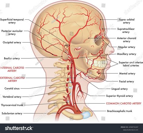
Table of Contents
Label the Arteries of the Head and Neck: A Comprehensive Guide
Understanding the arterial supply of the head and neck is crucial for medical professionals, students, and anyone interested in human anatomy. This intricate network of blood vessels delivers oxygen and nutrients to the brain, face, and neck, supporting their vital functions. This comprehensive guide will delve into the detailed anatomy of the arteries supplying this region, focusing on their origins, branches, and the areas they serve. We'll explore the major arteries, their clinical significance, and potential pathologies associated with them.
Major Arteries of the Head and Neck
The primary source of blood supply to the head and neck is the common carotid arteries, branching from the aortic arch. These bifurcate into the internal carotid arteries and external carotid arteries at the level of the upper border of the thyroid cartilage. Let's explore each in detail.
Internal Carotid Artery: Fueling the Brain
The internal carotid artery, unlike its external counterpart, does not directly contribute to the external structures of the head and neck. Its primary role is to supply the brain. It's a crucial vessel, and its blockage can lead to devastating consequences, such as stroke. It ascends through the neck, passing through the carotid canal of the temporal bone before entering the cranial cavity.
Branches of the Internal Carotid Artery:
While the internal carotid artery itself doesn't branch extensively in the neck, its intracranial branches are numerous and vital. These include:
-
Opthalmic Artery: Supplies the eye and its surrounding structures, including the orbit, eyelids, and lacrimal gland. Its branches are incredibly intricate and support the visual system's delicate functions. Blockages here can result in vision impairment.
-
Posterior Communicating Artery: Part of the Circle of Willis, a crucial anastomotic ring at the base of the brain that provides redundancy in cerebral blood supply. This artery connects the posterior cerebral artery to the internal carotid artery. Its role in maintaining consistent blood flow to the brain is critical.
-
Anterior Cerebral Artery: Supplies the medial surface of the frontal and parietal lobes of the brain. This is a significant artery for higher cognitive functions. Occlusion can lead to severe neurological deficits.
-
Middle Cerebral Artery: The largest branch of the internal carotid artery, supplying the lateral surface of the frontal, parietal, and temporal lobes. This artery is critically involved in motor function, speech, and sensory perception. It is frequently involved in ischemic strokes.
Clinical Significance of the Internal Carotid Artery: Given its role in supplying the brain, the internal carotid artery holds significant clinical importance. Stenosis (narrowing) or occlusion (blockage) can lead to ischemic strokes, causing devastating neurological deficits. Diagnosis often involves imaging techniques like carotid ultrasound and CT angiography.
External Carotid Artery: Nourishing the Face and Neck
The external carotid artery, in contrast to the internal carotid, branches extensively to supply the structures of the face, neck, and scalp. It has eight main branches, generally categorized into anterior, posterior, and medial groups.
Branches of the External Carotid Artery:
-
Anterior Branches:
-
Superior Thyroid Artery: Supplies the thyroid gland, larynx, and trachea. Its involvement in thyroid surgeries is significant.
-
Lingual Artery: Provides blood to the tongue, supplying the muscles of the tongue and its sensory structures.
-
Facial Artery: A prominent artery with multiple branches supplying the face, including the muscles of facial expression, lips, nose, and the submandibular region. This artery is frequently involved in surgical procedures related to facial reconstruction.
-
-
Posterior Branches:
-
Occipital Artery: Supplies the back of the scalp and the posterior neck muscles.
-
Posterior Auricular Artery: Provides blood to the posterior ear, mastoid process, and external acoustic meatus.
-
-
Medial Branches:
- Ascending Pharyngeal Artery: A small but crucial artery supplying the pharynx and the surrounding structures, including the soft palate.
-
Terminal Branches:
-
Superficial Temporal Artery: Supplies the temporal region of the scalp, the auricle, and the parotid gland. It is easily palpable in front of the ear and is a common site for temporal artery biopsies.
-
Maxillary Artery: A deep artery that travels through the infratemporal fossa, providing branches to the teeth, nasal cavity, meninges, and deeper structures of the face. It’s often involved in procedures related to dental surgery and treatment of nasal pathology.
-
Clinical Significance of the External Carotid Artery: The external carotid artery, due to its superficial course, is often accessible for palpation and examination. Its branches are important in surgical procedures involving the face, neck, and scalp. Injury to these vessels can lead to significant bleeding. Furthermore, understanding the branching pattern is crucial for interpreting imaging studies and planning surgical approaches.
Vertebral Artery: Supplying the Posterior Brain
While not directly a branch of the common carotid arteries, the vertebral arteries are critical in supplying blood to the brain. These arteries arise from the subclavian arteries and ascend through the transverse foramina of the cervical vertebrae, entering the skull through the foramen magnum. They merge to form the basilar artery, which plays a key role in the Circle of Willis.
Branches of the Vertebral and Basilar Arteries:
-
Vertebral Artery Branches: Before joining to form the basilar artery, the vertebral arteries supply blood to parts of the brainstem and cerebellum.
-
Basilar Artery Branches: The basilar artery gives off numerous branches that irrigate the brainstem, cerebellum, and occipital lobes. These include the pontine arteries, anterior inferior cerebellar arteries, superior cerebellar arteries, and the posterior cerebral arteries which are crucial for maintaining vital functions.
Clinical Significance of the Vertebral Artery: Occlusion or stenosis of the vertebral arteries can cause vertebral-basilar insufficiency, leading to symptoms such as dizziness, vertigo, diplopia (double vision), and ataxia (loss of coordination).
The Circle of Willis: A Redundant Blood Supply System
The Circle of Willis is a remarkable anastomotic ring of arteries at the base of the brain. This interconnected network of arteries—formed by the anterior and posterior communicating arteries, along with segments of the internal carotid and vertebral arteries—serves as a crucial backup system for cerebral blood flow. If one major artery is blocked, the Circle of Willis can help to reroute blood flow and minimize the impact of the blockage.
Clinical Correlations and Pathologies
Understanding the arterial supply of the head and neck is critical for diagnosing and managing various pathologies. Here are some important clinical considerations:
-
Stroke: Ischemic stroke, resulting from blockage of an artery, is a major cause of neurological deficits. Knowing the arterial distribution helps determine the affected brain regions and predict the neurological consequences.
-
Aneurysms: Bulges in artery walls can rupture, leading to life-threatening hemorrhagic strokes. The location of an aneurysm within the head and neck arterial system is crucial for surgical planning.
-
Trauma: Penetrating or blunt trauma to the head and neck can damage arteries, leading to significant bleeding.
-
Temporal Arteritis: Inflammation of the temporal arteries can cause headaches, visual disturbances, and jaw claudication.
Diagnostic Imaging Techniques
Various imaging techniques are used to visualize the arteries of the head and neck:
-
Carotid Ultrasound: A non-invasive technique to assess blood flow in the carotid arteries.
-
CT Angiography: A detailed imaging technique using contrast dye to visualize the arteries.
-
MRI Angiography: A similar technique to CT angiography but using magnetic resonance imaging.
-
Digital Subtraction Angiography (DSA): A more invasive technique that provides high-resolution images of blood vessels.
Conclusion
The arterial supply of the head and neck is a complex and intricate system crucial for supporting the vital functions of the brain, face, and neck. Understanding the origins, branches, and clinical significance of these arteries is fundamental to various medical disciplines. This comprehensive overview provided a thorough exploration of this critical anatomical area, enabling better comprehension of its complexities and clinical implications. Further study and exploration of this area will enhance understanding and lead to improved diagnostics and therapeutic strategies. The continued advancement in medical imaging allows for refined investigation, leading to even more nuanced insights into this critical system. This detailed understanding underpins the advancements in neurosurgery, vascular surgery, and other medical specialties relying on a precise knowledge of the head and neck vasculature.
Latest Posts
Latest Posts
-
How To Collect Urine Specimen From Foley Catheter
Mar 28, 2025
-
The Idea Of Spontaneous Generation Postulated That
Mar 28, 2025
-
How Many Movements Are In A Classical Concerto
Mar 28, 2025
-
What Is Necessary For Diffusion To Occur
Mar 28, 2025
-
Color Of Flame Of Calcium Chloride
Mar 28, 2025
Related Post
Thank you for visiting our website which covers about Label The Arteries Of The Head And Neck . We hope the information provided has been useful to you. Feel free to contact us if you have any questions or need further assistance. See you next time and don't miss to bookmark.
