Label The Microscopic Anatomy Of Spongy Bone
Muz Play
Mar 22, 2025 · 6 min read
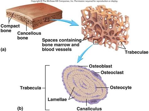
Table of Contents
- Label The Microscopic Anatomy Of Spongy Bone
- Table of Contents
- Labeling the Microscopic Anatomy of Spongy Bone: A Comprehensive Guide
- I. The Trabeculae: The Scaffolding of Spongy Bone
- II. Bone Marrow: The Life-Giving Center
- III. Osteocytes: The Bone Cells Within the Trabeculae
- IV. Other Cellular Components: Supporting the Bone Remodeling Process
- V. Blood Vessels and Nerves: The Supply Lines
- VI. Microscopic Appearance and Labeling Exercise
- VII. Clinical Significance: Understanding Diseases and Injuries
- VIII. Conclusion: A Foundation for Further Study
- Latest Posts
- Latest Posts
- Related Post
Labeling the Microscopic Anatomy of Spongy Bone: A Comprehensive Guide
Spongy bone, also known as cancellous bone or trabecular bone, is a type of bone tissue that forms the interior of most bones. Unlike compact bone, which is dense and solid, spongy bone is characterized by its porous structure, consisting of a network of interconnected bony spicules called trabeculae. Understanding the microscopic anatomy of spongy bone is crucial for grasping its function and its role in the overall skeletal system. This detailed guide will walk you through the key components, providing a comprehensive understanding for labeling purposes and beyond.
I. The Trabeculae: The Scaffolding of Spongy Bone
The most striking feature of spongy bone under a microscope is the trabeculae. These are thin, interconnected bony plates or struts that create a three-dimensional lattice-like structure. The spaces between the trabeculae are filled with bone marrow, a crucial component involved in hematopoiesis (blood cell production).
-
Structure of Trabeculae: Trabeculae are not randomly arranged. Their orientation is highly organized, reflecting the lines of stress placed on the bone. They are composed of lamellae, similar to those found in compact bone, but these lamellae are less regularly arranged. They often lack the concentric rings seen in osteons (Haversian systems) of compact bone.
-
Composition of Trabeculae: Trabeculae are primarily composed of osteocytes housed within lacunae. These lacunae are interconnected by canaliculi, tiny channels that allow for communication and nutrient exchange between osteocytes. The trabeculae also contain a network of blood vessels that run through the spaces between the bony struts, providing nutrients to the bone cells and transporting waste products away.
II. Bone Marrow: The Life-Giving Center
The spaces between the trabeculae are filled with bone marrow. There are two main types of bone marrow:
-
Red Bone Marrow (Myeloid Tissue): This is the hematopoietic tissue responsible for the production of red blood cells, white blood cells, and platelets. It's rich in blood vessels and hematopoietic stem cells. Red marrow is particularly abundant in the spongy bone of flat bones like the sternum, ribs, and hip bones, as well as in the epiphyses (ends) of long bones. Under the microscope, red marrow appears as a cellular, highly vascularized tissue.
-
Yellow Bone Marrow: As individuals age, much of the red bone marrow is replaced by yellow bone marrow, which is primarily composed of adipose tissue (fat). While less active in hematopoiesis, yellow bone marrow can revert back to red marrow under certain conditions, such as significant blood loss.
III. Osteocytes: The Bone Cells Within the Trabeculae
Osteocytes are mature bone cells that reside within lacunae within the trabeculae. They are responsible for maintaining the bone matrix and sensing mechanical stress on the bone. Their interconnectedness through canaliculi is vital for communication and nutrient exchange.
-
Lacunae: These are small, hollow spaces within the bone matrix where the osteocytes reside. They appear as dark, empty spaces under the microscope.
-
Canaliculi: These are minute canals that radiate from the lacunae, connecting them to each other and to the blood vessels. They allow for the passage of nutrients and waste products between osteocytes.
IV. Other Cellular Components: Supporting the Bone Remodeling Process
While osteocytes are the dominant cell type within spongy bone, other cell types play crucial roles in bone remodeling and maintenance:
-
Osteoblasts: These are bone-forming cells responsible for synthesizing and depositing new bone matrix (osteoid). They are found on the surfaces of trabeculae, actively building new bone.
-
Osteoclasts: These are large, multinucleated cells responsible for bone resorption, the process of breaking down bone tissue. They are involved in bone remodeling and calcium regulation. They are less numerous but equally critical in maintaining bone health.
-
Bone Lining Cells: These cells cover bone surfaces that are not actively undergoing remodeling. They play a role in maintaining the integrity of the bone surface.
V. Blood Vessels and Nerves: The Supply Lines
Spongy bone is richly vascularized, with blood vessels running through the spaces between the trabeculae and supplying the bone marrow and osteocytes. The arrangement of these blood vessels is irregular compared to the highly organized systems found in compact bone. Nerves accompany the blood vessels, providing sensory innervation to the bone.
VI. Microscopic Appearance and Labeling Exercise
When observing spongy bone under a microscope (typically with a light microscope and potentially specialized staining techniques), you'll see the following:
-
Trabeculae: The prominent network of interconnected bony struts. These will appear as a three-dimensional latticework, stained differently depending on the staining method used.
-
Lacunae: Small, dark spaces within the trabeculae, housing the osteocytes.
-
Canaliculi: Fine, radiating lines extending from the lacunae, connecting them to each other and blood vessels. These might be challenging to clearly visualize without specialized staining.
-
Bone Marrow: The spaces between trabeculae filled with either red marrow (cellular and vascularized) or yellow marrow (adipose tissue). The appearance will vary based on age and location.
-
Osteocytes (with staining): Using appropriate stains, osteocytes might be visible as small, dark structures within the lacunae.
-
Osteoblasts (with staining): These would appear as cuboidal cells on the surface of trabeculae, actively synthesizing bone matrix.
-
Osteoclasts (with staining): These large, multinucleated cells can be identified on the surfaces of trabeculae where bone resorption is taking place.
-
Blood Vessels (with staining): These appear as dark, round or elongated structures within the marrow spaces and running along the trabeculae.
Labeling Practice: To effectively label a microscopic image of spongy bone, you should be able to identify and label each of the above components. Practice drawing diagrams and labeling the key structures will significantly improve your understanding.
VII. Clinical Significance: Understanding Diseases and Injuries
Understanding the microscopic anatomy of spongy bone is crucial for diagnosing and treating a range of clinical conditions. Here are a few examples:
-
Osteoporosis: This condition is characterized by reduced bone mass and density, leading to increased fragility and fracture risk. Microscopic examination of spongy bone in osteoporosis reveals thinner and less interconnected trabeculae.
-
Bone Tumors: Both benign and malignant bone tumors can affect spongy bone. Microscopic analysis is vital in determining the type and extent of the tumor.
-
Bone Fractures: Understanding the structural properties of spongy bone is critical in assessing the severity and treatment of bone fractures, especially those affecting the cancellous portion of bones.
-
Blood Disorders: Since spongy bone contains red bone marrow, abnormalities in blood cell production can often be identified through microscopic examination of bone marrow samples.
VIII. Conclusion: A Foundation for Further Study
This comprehensive guide provides a strong foundation for understanding and labeling the microscopic anatomy of spongy bone. By mastering the identification of key structures like trabeculae, bone marrow, osteocytes, and other cellular components, you'll develop a deeper appreciation of this crucial tissue's role in supporting the body's skeletal framework and hematopoietic functions. Further research into specific staining techniques and the use of advanced microscopy will enhance your ability to visualize and understand the intricate details of spongy bone's microscopic architecture. Remember that practice is key, so utilize diagrams, microscopic images, and labeling exercises to solidify your knowledge.
Latest Posts
Latest Posts
-
What Is The Relationship Between Temperature And Pressure
Mar 25, 2025
-
Is Toxicity A Physical Or Chemical Property
Mar 25, 2025
-
Plasma Membrane In A Plant Cell
Mar 25, 2025
-
What Are The P Block Elements
Mar 25, 2025
-
Design Primers For Site Directed Mutagenesis
Mar 25, 2025
Related Post
Thank you for visiting our website which covers about Label The Microscopic Anatomy Of Spongy Bone . We hope the information provided has been useful to you. Feel free to contact us if you have any questions or need further assistance. See you next time and don't miss to bookmark.
