Label The Structures Of The Abdominopelvic Cavity
Muz Play
Mar 22, 2025 · 7 min read
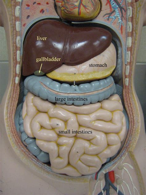
Table of Contents
Labeling the Structures of the Abdominopelvic Cavity: A Comprehensive Guide
The abdominopelvic cavity, a vast and complex space, houses a multitude of vital organs. Understanding its anatomy is crucial for anyone in the medical field, as well as for students of anatomy and physiology. This comprehensive guide will delve into the detailed labeling of the structures within this cavity, providing a robust foundation for your knowledge. We will explore the various regions, organs, and their specific locations, equipping you with the skills to accurately identify and label each component.
Divisions of the Abdominopelvic Cavity
Before diving into the specific structures, it's important to understand how the abdominopelvic cavity is divided. This segmentation helps in organizing and understanding the location of various organs. The cavity is broadly divided into two main sections:
1. Abdominal Cavity:
The superior portion, containing the majority of the digestive organs. It's further subdivided for easier identification and study using several methods:
-
Quadrants: The simplest division involves four quadrants formed by two imaginary lines intersecting at the umbilicus (navel): the right upper quadrant (RUQ), left upper quadrant (LUQ), right lower quadrant (RLQ), and left lower quadrant (LLQ). This is commonly used in clinical settings for quick localization of pain or abnormalities.
-
Nine Regions: A more detailed division uses nine regions defined by four imaginary lines: two midclavicular lines (vertical lines passing through the midpoints of the clavicles) and two horizontal lines (the subcostal line passing just below the ribs and the transtubercular line passing across the superior aspects of the hip bones). This system provides more precise anatomical location:
- Right Hypochondriac Region: Located superior and lateral to the epigastric region. Houses the right lobe of the liver, gallbladder, and parts of the right kidney and colon.
- Epigastric Region: The superior midline region, overlying the stomach and part of the liver.
- Left Hypochondriac Region: Superior and lateral to the epigastric region. Contains the left lobe of the liver, stomach, spleen, and parts of the left kidney and colon.
- Right Lumbar Region: Lateral to the umbilical region. Contains the ascending colon and parts of the right kidney and small intestine.
- Umbilical Region: The central region surrounding the umbilicus. Contains parts of the small intestine, transverse colon, and inferior vena cava.
- Left Lumbar Region: Lateral to the umbilical region. Contains the descending colon and parts of the left kidney and small intestine.
- Right Iliac (Inguinal) Region: Inferior and lateral to the umbilical region. Contains the cecum, appendix, and parts of the small intestine.
- Hypogastric (Pubic) Region: The inferior midline region. Contains the urinary bladder, parts of the sigmoid colon, and the uterus (in females).
- Left Iliac (Inguinal) Region: Inferior and lateral to the umbilical region. Contains the sigmoid colon and parts of the small intestine.
2. Pelvic Cavity:
The inferior portion, situated within the bony pelvis. It houses organs related to reproduction, urination, and the terminal part of the digestive system. Key structures include:
- Urinary Bladder: A muscular sac storing urine.
- Rectum: The terminal portion of the large intestine, storing feces before elimination.
- Reproductive Organs: These vary significantly between males and females. In females, this includes the uterus, fallopian tubes, and ovaries. In males, this includes the prostate gland, seminal vesicles, and parts of the vas deferens.
Detailed Labeling of Key Abdominal Organs
Now let's delve into the detailed labeling of specific organs within the abdominopelvic cavity, keeping in mind the anatomical divisions discussed above. Remember, the precise location of some organs can vary slightly between individuals.
Organs of the Digestive System:
- Stomach: A J-shaped organ located in the left upper quadrant (LUQ) and epigastric region, responsible for food storage and initial digestion.
- Small Intestine: A long, coiled tube extending from the stomach to the large intestine. It's divided into three parts: the duodenum (mostly in the RUQ), jejunum (mostly in the LUQ and umbilical region), and ileum (mostly in the RLQ and LLQ). The small intestine is the primary site of nutrient absorption.
- Large Intestine: A wider tube extending from the small intestine to the anus. It consists of the cecum (RLQ), ascending colon (RUQ), transverse colon (spanning across the umbilical region), descending colon (LUQ), sigmoid colon (LLQ), rectum (pelvic cavity), and anus. The large intestine absorbs water and electrolytes, forming and storing feces.
- Liver: The largest gland in the body, located primarily in the right upper quadrant (RUQ), extending slightly into the epigastric region. It performs a multitude of functions, including bile production, detoxification, and nutrient storage.
- Gallbladder: A small sac located beneath the liver (RUQ), storing and concentrating bile produced by the liver.
- Pancreas: A gland located behind the stomach (LUQ), producing digestive enzymes and hormones like insulin.
- Spleen: An organ located in the left upper quadrant (LUQ), playing a vital role in the immune system and filtering blood.
- Appendix: A small, finger-like projection from the cecum (RLQ), often associated with appendicitis.
Organs of the Urinary System:
- Kidneys: Two bean-shaped organs located retroperitoneally (behind the peritoneum) on either side of the vertebral column. They filter blood and produce urine. Parts extend into the lumbar regions.
- Ureters: Two tubes carrying urine from the kidneys to the urinary bladder.
- Urinary Bladder: A muscular sac in the pelvic cavity storing urine before elimination.
- Urethra: A tube carrying urine from the bladder to the outside of the body.
Organs of the Reproductive System:
The location and presence of reproductive organs greatly vary between sexes.
Female Reproductive Organs:
- Ovaries: Two almond-shaped organs located in the pelvic cavity, producing eggs and hormones.
- Fallopian Tubes: Two tubes connecting the ovaries to the uterus, transporting eggs.
- Uterus: A muscular organ in the pelvic cavity, housing the developing fetus during pregnancy.
- Vagina: A canal connecting the uterus to the outside of the body.
Male Reproductive Organs:
- Testes: Two oval-shaped organs located within the scrotum, producing sperm and testosterone.
- Epididymis: A coiled tube atop each testis, storing and maturing sperm.
- Vas Deferens: A tube carrying sperm from the epididymis to the urethra.
- Seminal Vesicles: Glands producing seminal fluid.
- Prostate Gland: A gland surrounding the urethra, producing seminal fluid.
Importance of Accurate Labeling
Accurate labeling of the abdominopelvic cavity structures is crucial for several reasons:
- Medical Diagnosis: Precise labeling allows for accurate communication between medical professionals regarding the location of abnormalities, injuries, or diseases.
- Surgical Procedures: Surgeons rely heavily on anatomical knowledge to navigate the abdominopelvic cavity safely and effectively during operations.
- Medical Imaging Interpretation: Correct labeling of structures is essential for interpreting medical images like X-rays, CT scans, and MRIs.
- Anatomical Understanding: Accurate labeling fosters a deeper understanding of the complex interplay between organs and systems within the abdominopelvic cavity.
Utilizing Resources for Learning
Several resources can assist in mastering the labeling of abdominopelvic cavity structures:
- Anatomical Models: Three-dimensional models provide a tactile learning experience, allowing for hands-on exploration of the organs and their relationships.
- Anatomical Atlases: Detailed atlases provide high-quality images and descriptions of the various structures within the cavity.
- Interactive Anatomy Software: Computer programs offer interactive exploration of the abdominopelvic cavity, allowing users to zoom in, rotate, and dissect virtual organs.
- Flashcards and Quizzes: Utilizing flashcards and online quizzes provides a effective way to test and reinforce your knowledge.
Conclusion
Mastering the labeling of structures within the abdominopelvic cavity requires dedicated study and practice. By utilizing the information presented here and various learning resources, you can build a solid foundation of anatomical knowledge. This knowledge is not only essential for medical professionals but also incredibly valuable for anyone seeking a comprehensive understanding of human anatomy. Remember to use a combination of learning methods to reinforce your understanding and ensure you can accurately label all the key organs and their relative positions within the abdominopelvic cavity. Through consistent effort and practice, you can achieve a proficient level of understanding and labeling accuracy. Continue to challenge yourself with quizzes and practical application to solidify your skills and contribute to a strong understanding of this crucial anatomical region.
Latest Posts
Latest Posts
-
Determine Whether The Graph Is The Graph Of A Function
Mar 22, 2025
-
Lewis Structure Practice Worksheet And Answers
Mar 22, 2025
-
The Cell The Basic Unit Of Life
Mar 22, 2025
-
Power Series Solution Of Ordinary Differential Equations
Mar 22, 2025
-
How Many Electrons Does Li2 Have
Mar 22, 2025
Related Post
Thank you for visiting our website which covers about Label The Structures Of The Abdominopelvic Cavity . We hope the information provided has been useful to you. Feel free to contact us if you have any questions or need further assistance. See you next time and don't miss to bookmark.
