Microscopic Anatomy Of Skeletal Muscle Fiber
Muz Play
Mar 15, 2025 · 6 min read
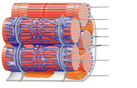
Table of Contents
Microscopic Anatomy of Skeletal Muscle Fiber: A Deep Dive
Skeletal muscle, the powerhouse of voluntary movement, is a marvel of biological engineering. Understanding its microscopic anatomy is crucial to comprehending how we move, how we exert force, and how muscle diseases manifest. This article delves deep into the fascinating microscopic world of the skeletal muscle fiber, exploring its intricate structure and the components that contribute to its remarkable functionality.
The Muscle Fiber: A Functional Unit
The skeletal muscle fiber, also known as a muscle cell or myofiber, is the basic functional unit of skeletal muscle. These long, cylindrical cells are multinucleated, meaning they contain multiple nuclei located just beneath the sarcolemma, the muscle fiber's plasma membrane. This multinucleated nature is a result of the fusion of many myoblasts during development. The size of muscle fibers varies considerably, depending on the muscle's function and the individual's training status. However, they generally range from 10 to 100 micrometers in diameter and can extend the entire length of the muscle.
Sarcolemma and Transverse Tubules (T-tubules)
The sarcolemma plays a crucial role in transmitting electrical signals that initiate muscle contraction. It's not just a passive barrier; it actively participates in the excitation-contraction coupling process. Deep invaginations of the sarcolemma, known as transverse tubules or T-tubules, penetrate the fiber at the junctions between the A and I bands (explained further below). These T-tubules are crucial for rapidly transmitting the action potential from the surface of the muscle fiber to the interior, ensuring that all parts of the fiber contract simultaneously. The close association of T-tubules with the sarcoplasmic reticulum (SR) is critical for the efficient release of calcium ions, the trigger for muscle contraction.
Sarcoplasmic Reticulum (SR) and Calcium Ion Regulation
The sarcoplasmic reticulum (SR) is a specialized endoplasmic reticulum found within muscle fibers. It forms a network of interconnected sacs and tubules surrounding each myofibril. The SR's primary function is to store and release calcium ions (Ca²⁺). When an action potential arrives via the T-tubules, it triggers the release of Ca²⁺ from the SR into the sarcoplasm (the cytoplasm of the muscle fiber). This sudden increase in cytosolic Ca²⁺ concentration initiates the interaction between actin and myosin filaments, leading to muscle contraction. The precise regulation of Ca²⁺ release and reuptake by the SR is essential for controlling the duration and strength of muscle contraction. Specialized proteins within the SR, such as the ryanodine receptor (RyR) and the sarco/endoplasmic reticulum Ca²⁺-ATPase (SERCA), play key roles in this finely tuned process.
Myofibrils: The Contractile Machinery
Within each muscle fiber, numerous myofibrils run parallel to the long axis of the cell. These cylindrical structures are the actual contractile elements of the muscle fiber, responsible for generating the force of muscle contraction. Myofibrils are highly organized bundles of protein filaments, primarily actin (thin filaments) and myosin (thick filaments), arranged in repeating units called sarcomeres.
Sarcomeres: The Basic Contractile Unit
The sarcomere is the fundamental unit of muscle contraction. These highly organized structures extend from one Z-line (or Z-disc) to the next. The Z-line is a dense protein structure that anchors the thin filaments. Key structural components within the sarcomere include:
- A band: The anisotropic band, appearing dark under a microscope, contains the entire length of the thick myosin filaments and the overlapping regions with thin actin filaments.
- I band: The isotropic band, appearing light under a microscope, contains only thin actin filaments and extends from the A band of one sarcomere to the A band of the adjacent sarcomere. The Z-line is located in the center of the I band.
- H zone: A lighter area within the A band where only thick myosin filaments are present, without overlap with thin actin filaments. The M line runs through the center of the H zone, providing structural support to the thick filaments.
- M line: A protein structure in the center of the sarcomere that anchors the thick filaments and helps maintain their alignment.
Actin and Myosin Filaments: The Molecular Players
The sliding filament theory explains muscle contraction as the result of the relative movement of actin and myosin filaments within the sarcomere. This movement is driven by the interaction between myosin heads and actin filaments.
- Myosin: Myosin filaments are composed of numerous myosin molecules, each with a globular head and a tail region. The myosin heads possess ATPase activity, allowing them to bind to actin, hydrolyze ATP, and generate the force for sliding.
- Actin: Actin filaments are primarily composed of actin monomers polymerized into a double helix. Troponin and tropomyosin are regulatory proteins associated with actin filaments. Tropomyosin covers the myosin-binding sites on actin in the relaxed state, preventing interaction between actin and myosin. Troponin, a complex of three proteins, binds to tropomyosin and calcium ions. The binding of calcium to troponin causes a conformational change, moving tropomyosin and exposing the myosin-binding sites on actin, allowing for the cross-bridge cycle to begin.
The Cross-Bridge Cycle: The Mechanism of Contraction
The cross-bridge cycle describes the cyclical interaction between myosin heads and actin filaments, leading to muscle contraction. This process is driven by ATP hydrolysis:
- Attachment: A myosin head, in its high-energy configuration (bound to ADP and Pi), binds to an exposed myosin-binding site on actin.
- Power Stroke: The myosin head pivots, causing the actin filament to slide past the myosin filament. ADP and Pi are released during this power stroke.
- Detachment: A new ATP molecule binds to the myosin head, causing it to detach from actin.
- Cocking: ATP is hydrolyzed to ADP and Pi, returning the myosin head to its high-energy configuration, ready to bind to another actin molecule and repeat the cycle.
Other Important Components
Beyond the sarcomere, other components contribute to the overall structure and function of the muscle fiber:
- Mitochondria: Abundant mitochondria are crucial for supplying the ATP required for muscle contraction. Their high density within muscle fibers reflects the energy demands of this process.
- Glycogen granules: These store glycogen, a readily available source of glucose that can be broken down to provide energy for contraction.
- Myoglobin: This oxygen-binding protein stores oxygen within muscle fibers, facilitating aerobic respiration and delaying fatigue.
- Connective Tissue: Muscle fibers are bundled together by connective tissue, providing structural support and transmitting the force of contraction to the tendons.
Implications for Health and Disease
Understanding the microscopic anatomy of skeletal muscle fiber is essential for diagnosing and treating various muscle disorders. Disruptions in any of the components discussed above can lead to impaired muscle function. For example:
- Muscular dystrophies: These genetic diseases affect the structure of muscle proteins, leading to muscle weakness and degeneration.
- Myasthenia gravis: An autoimmune disease that affects the neuromuscular junction, impairing the transmission of signals from nerves to muscles.
- Muscle cramps: These painful spasms can result from electrolyte imbalances or excessive muscle activity.
- Age-related muscle loss (sarcopenia): A gradual decline in muscle mass and function with age, often associated with decreased protein synthesis and increased muscle fiber atrophy.
Conclusion
The microscopic anatomy of skeletal muscle fiber is a complex and fascinating subject, reflecting the intricate mechanisms responsible for our ability to move. From the multinucleated structure of the muscle fiber to the precise orchestration of the cross-bridge cycle within the sarcomere, each component plays a critical role in muscle function. A thorough understanding of this anatomy is crucial not only for appreciating the beauty of biological systems but also for addressing the challenges of various muscle disorders and developing effective therapeutic strategies. Further research into the intricacies of skeletal muscle will continue to unravel the mysteries of this remarkable tissue and provide valuable insights into maintaining healthy muscle function throughout life.
Latest Posts
Latest Posts
-
Can Starch Pass Through Dialysis Tubing
Mar 15, 2025
-
How Do You Find Average Acceleration
Mar 15, 2025
-
Is O Or S More Electronegative
Mar 15, 2025
-
If An Atom Gains An Electron It Becomes A
Mar 15, 2025
-
How To Determine Shape Of A Molecule
Mar 15, 2025
Related Post
Thank you for visiting our website which covers about Microscopic Anatomy Of Skeletal Muscle Fiber . We hope the information provided has been useful to you. Feel free to contact us if you have any questions or need further assistance. See you next time and don't miss to bookmark.
