Shaft Of Long Bone Is Called
Muz Play
Mar 20, 2025 · 6 min read
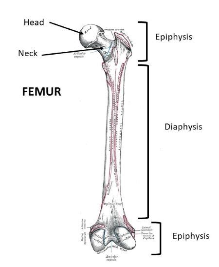
Table of Contents
The Diaphysis: Understanding the Shaft of a Long Bone
The shaft of a long bone, also known as the diaphysis, is a crucial part of the skeletal system, contributing significantly to the overall structure, function, and growth of long bones. Understanding its anatomy, composition, and role in the body is essential for anyone studying anatomy, physiology, or related fields. This comprehensive guide delves into the diaphysis, exploring its characteristics, development, clinical significance, and related concepts.
What is the Diaphysis?
The diaphysis is the elongated, cylindrical shaft that forms the main portion of a long bone. It's located between the two ends of the bone, the epiphyses. Think of it as the long, straight part of a bone like your femur (thigh bone) or humerus (upper arm bone). This central section provides structural support and leverage for movement. Its primary function is to bear weight and withstand stress during activities like walking, running, and lifting.
Key Characteristics of the Diaphysis:
- Shape: Generally cylindrical, but can exhibit slight variations depending on the specific bone.
- Structure: Primarily composed of compact bone, a dense and strong type of bone tissue. This provides maximum strength with minimal weight.
- Cavity: Contains the medullary cavity, a hollow space filled with bone marrow. In adults, this marrow is primarily yellow marrow (fatty tissue), while in children, it's primarily red marrow (responsible for blood cell production).
- Covering: Surrounded by a tough, fibrous membrane called the periosteum, which is crucial for bone growth, repair, and nutrient supply. The periosteum is richly supplied with blood vessels and nerves.
Composition of the Diaphysis: A Closer Look
The diaphysis's compact bone structure is highly organized. It consists of tightly packed osteons, also known as Haversian systems. Each osteon is a cylindrical unit composed of concentric lamellae (layers) of bone tissue surrounding a central Haversian canal. These canals contain blood vessels and nerves that nourish the bone cells (osteocytes). The osteocytes reside within lacunae (small cavities) within the lamellae, connected to each other and the Haversian canals via canaliculi (tiny channels). This intricate network ensures efficient nutrient delivery and waste removal.
The Role of Compact Bone:
The dense nature of compact bone in the diaphysis makes it exceptionally strong and resistant to bending and fracturing. This is critical for supporting the body's weight and facilitating movement. The tightly packed structure minimizes the risk of stress fractures and other injuries during physical activity.
Development of the Diaphysis: From Cartilage to Bone
Long bone development is a fascinating process involving endochondral ossification, where cartilage is gradually replaced by bone tissue. The diaphysis is one of the first parts of the long bone to undergo ossification. This process begins with a cartilaginous model of the bone, which serves as a template for bone formation. Ossification centers appear in the diaphysis, and bone tissue gradually replaces the cartilage.
Stages of Diaphyseal Ossification:
- Formation of the Cartilage Model: A hyaline cartilage model forms the shape of the future long bone.
- Appearance of the Primary Ossification Center: Blood vessels invade the cartilage model, bringing osteoblasts (bone-forming cells). Osteoblasts begin to deposit bone matrix within the diaphysis.
- Bone Collar Formation: A layer of compact bone (the bone collar) forms around the diaphysis.
- Cavitation: The interior of the diaphysis begins to break down, forming the medullary cavity.
- Secondary Ossification Centers: These appear later in the epiphyses (the ends of the long bones).
- Growth and Maturation: The diaphysis elongates as the cartilage continues to grow and is replaced by bone. Eventually, the epiphyseal plates (growth plates) close, marking the end of longitudinal bone growth.
Clinical Significance of the Diaphysis: Fractures and More
The diaphysis, due to its position and function, is susceptible to injury, especially fractures. Diaphyseal fractures are common and can result from a variety of mechanisms, including high-impact trauma, falls, and repetitive stress. The severity of a diaphyseal fracture can vary widely, ranging from simple hairline cracks to complex comminuted fractures (where the bone is shattered into multiple pieces).
Common Types of Diaphyseal Fractures:
- Transverse fractures: The fracture line runs perpendicular to the long axis of the bone.
- Oblique fractures: The fracture line runs at an angle to the long axis of the bone.
- Spiral fractures: The fracture line spirals around the bone, often indicating a twisting injury.
- Comminuted fractures: The bone is broken into multiple fragments.
Treatment of Diaphyseal Fractures:
Treatment depends on the severity of the fracture and the patient's overall health. Options include:
- Casting or splinting: Used for less severe fractures to immobilize the bone and allow it to heal naturally.
- Surgery: May be necessary for more complex fractures, involving techniques like open reduction and internal fixation (ORIF), where plates, screws, or rods are used to stabilize the bone.
The Diaphysis and Bone Marrow: A Vital Partnership
The medullary cavity within the diaphysis houses bone marrow. In children, the bone marrow is primarily red marrow, responsible for hematopoiesis (blood cell production). As individuals age, much of the red marrow is replaced by yellow marrow, which is mainly adipose tissue (fat). However, some red marrow persists in specific locations even in adults.
The Significance of Bone Marrow:
Bone marrow plays a vital role in the body's overall health:
- Hematopoiesis: The production of red blood cells, white blood cells, and platelets, which are essential for oxygen transport, immune defense, and blood clotting.
- Immune System Support: Bone marrow contains various immune cells, contributing to the body's defense against infections.
- Fat Storage: Yellow marrow serves as a reservoir for energy storage.
The Periosteum: Protecting and Nourishing the Diaphysis
The periosteum is a crucial structure covering the diaphysis and other bones. It's a fibrous membrane consisting of two layers: the outer fibrous layer and the inner osteogenic layer. The outer layer provides protection and attachment points for tendons and ligaments. The inner layer contains osteoblasts and osteoprogenitor cells, responsible for bone growth and repair.
Functions of the Periosteum:
- Bone Growth and Repair: The periosteum plays a vital role in bone remodeling and fracture healing.
- Nutrient Supply: It provides blood vessels that nourish the underlying bone tissue.
- Protection: It acts as a protective barrier, safeguarding the bone from injury.
- Attachment: Provides points of attachment for tendons and ligaments.
Diaphysis and Related Concepts: Epiphysis and Metaphysis
Understanding the diaphysis requires understanding its relationship with other parts of the long bone:
- Epiphysis: The ends of the long bone, composed primarily of spongy bone and covered with articular cartilage. They provide articulating surfaces for joints.
- Metaphysis: The transitional region between the diaphysis and epiphysis, containing the epiphyseal plate (growth plate) in children and adolescents. This is the area where longitudinal bone growth occurs. Once growth is complete, the epiphyseal plate closes, leaving the epiphyseal line.
Conclusion: The Diaphysis – A Foundation of Skeletal Health
The diaphysis, the shaft of a long bone, is a critical component of the skeletal system. Its robust structure, primarily composed of compact bone, provides the strength and support needed for weight-bearing and movement. Understanding its development, composition, and clinical significance is vital for comprehending the overall function and health of the human skeleton. Furthermore, its close relationship with the bone marrow and periosteum highlights its critical role in various bodily functions, emphasizing the interconnectedness of different skeletal elements in maintaining overall health. Future research may lead to further advancements in understanding and treating diaphyseal injuries and related conditions, ensuring optimal skeletal health and function.
Latest Posts
Latest Posts
-
What Is A Mixed Melting Point
Mar 20, 2025
-
How Do Hydrogen Ions Flow Through Atp Synthase
Mar 20, 2025
-
How To Draw A 3d Vector
Mar 20, 2025
-
What Is It Called When A Volcano Collapses On Itself
Mar 20, 2025
-
Difference Between Reference And Thematic Maps
Mar 20, 2025
Related Post
Thank you for visiting our website which covers about Shaft Of Long Bone Is Called . We hope the information provided has been useful to you. Feel free to contact us if you have any questions or need further assistance. See you next time and don't miss to bookmark.
