Subdivisions Of The Ventral Body Cavity
Muz Play
Apr 04, 2025 · 7 min read

Table of Contents
Subdivisions of the Ventral Body Cavity: A Deep Dive into Thoracic and Abdominopelvic Regions
The human body is a marvel of intricate organization, and understanding its structure is crucial for comprehending its functions. A key aspect of this understanding involves the body cavities, spaces that house and protect vital organs. This article delves deep into the subdivisions of the ventral body cavity, a significant space encompassing the thoracic and abdominopelvic cavities. We will explore the individual components, their contents, and their clinical significance.
The Ventral Body Cavity: A Protective Shell
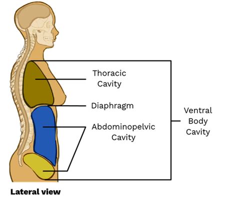
The ventral body cavity, also known as the coelom, is a large, fluid-filled space located on the anterior side of the body. Unlike the dorsal cavity (containing the cranial and vertebral cavities), the ventral cavity is responsible for housing the majority of the body’s viscera – the internal organs. Its primary function is protection; its fluid cushion protects these delicate organs from external shocks and impacts. The ventral cavity is further subdivided into two main cavities: the thoracic cavity and the abdominopelvic cavity. These are separated by the diaphragm, a dome-shaped muscle crucial for breathing.
1. The Thoracic Cavity: Heart, Lungs, and More
The thoracic cavity, located superior to the diaphragm, is a relatively closed space surrounded by the rib cage, sternum, and vertebral column. Its primary role is to protect the heart and lungs, but it also houses other vital structures. The thoracic cavity is further subdivided into three smaller cavities:
1.1. Pleural Cavities (x2): Home to the Lungs
There are two pleural cavities, one surrounding each lung. Each pleural cavity is a potential space, meaning it's normally only a thin layer of serous fluid, not an actual air-filled space. This serous fluid, secreted by the pleura (a thin, double-layered membrane), acts as a lubricant, minimizing friction during breathing. The visceral pleura covers the lungs directly, while the parietal pleura lines the inner surface of the thoracic cavity. The space between these layers is the pleural cavity itself. Inflammation of this pleural cavity is known as pleurisy, characterized by painful breathing.
Clinical Significance: Conditions like pneumothorax (collapsed lung) and pleural effusion (fluid buildup in the pleural space) directly impact the functionality of the pleural cavities. Understanding the anatomy of this space is vital for diagnosis and treatment.
1.2. Pericardial Cavity: Protecting the Heart
The pericardial cavity, located in the mediastinum, surrounds the heart. Similar to the pleural cavities, it is a potential space containing a small amount of serous fluid to reduce friction during heart contractions. The pericardium, a double-layered serous membrane, encloses the heart; the visceral pericardium adheres to the heart’s surface, while the parietal pericardium lines the pericardial sac. The space between these layers is the pericardial cavity.
Clinical Significance: Pericarditis (inflammation of the pericardium) and cardiac tamponade (fluid buildup compressing the heart) are serious conditions affecting the pericardial cavity, highlighting its importance in maintaining cardiac function.
1.3. Mediastinum: The Thoracic Cavity's Central Compartment
The mediastinum is the central compartment of the thoracic cavity, located between the two pleural cavities. It is not a true cavity, but rather a broad region containing several vital structures, including:
- The heart: Within the pericardial cavity.
- The great vessels: Major blood vessels like the aorta, vena cavae, and pulmonary arteries and veins.
- The trachea: The windpipe, transporting air to and from the lungs.
- The esophagus: The food pipe, transporting food from the mouth to the stomach.
- The thymus gland: An important part of the immune system, especially during childhood.
- Lymph nodes: Part of the body's lymphatic system.
Clinical Significance: Tumors, infections, and other conditions affecting the mediastinum can have widespread consequences due to the presence of so many critical structures.
2. The Abdominopelvic Cavity: Digestion, Excretion, and Reproduction
The abdominopelvic cavity, located inferior to the diaphragm, is a large cavity divided into two parts: the abdominal cavity and the pelvic cavity.
2.1. Abdominal Cavity: Housing Major Digestive Organs
The abdominal cavity contains the majority of the digestive organs, including the stomach, intestines (small and large), liver, gallbladder, pancreas, spleen, and kidneys. These organs are not directly exposed to the abdominal cavity; instead, they're enveloped by a serous membrane called the peritoneum.
The peritoneum is a double-layered membrane:
- Parietal peritoneum: Lines the inner surface of the abdominal wall.
- Visceral peritoneum: Covers the surfaces of the abdominal organs.
The space between the visceral and parietal peritoneum is called the peritoneal cavity, containing a small amount of serous fluid. Some organs are retroperitoneal, meaning they lie posterior to the peritoneum (e.g., kidneys, pancreas).
Clinical Significance: Conditions like peritonitis (inflammation of the peritoneum), appendicitis (inflammation of the appendix), and various gastrointestinal disorders directly involve structures within the abdominal cavity.
2.2. Pelvic Cavity: Reproductive and Urinary Organs
The pelvic cavity is the inferior part of the abdominopelvic cavity, enclosed by the bony pelvis. It houses the urinary bladder, rectum, internal reproductive organs (uterus, ovaries, fallopian tubes in females; prostate gland, seminal vesicles in males), and parts of the large intestine.
Clinical Significance: Conditions affecting the reproductive and urinary systems, such as urinary tract infections (UTIs), endometriosis, and prostate cancer, directly impact the pelvic cavity.
Abdominopelvic Regions and Quadrants: A Practical Approach
To facilitate the study and localization of abdominal and pelvic organs, the abdominopelvic cavity is often divided into regions or quadrants.
Abdominopelvic Regions (Nine): A More Detailed Subdivision
This method divides the abdominopelvic cavity into nine regions using four imaginary lines: two horizontal and two vertical. These regions are:
- Right hypochondriac region: Located superior and lateral to the umbilical region. Contains parts of the liver, gallbladder, and right kidney.
- Epigastric region: Located superior to the umbilical region. Contains the stomach, liver, and part of the pancreas.
- Left hypochondriac region: Located superior and lateral to the umbilical region. Contains parts of the stomach, spleen, and left kidney.
- Right lumbar region: Located lateral to the umbilical region. Contains parts of the ascending colon and right kidney.
- Umbilical region: Located in the center of the abdomen. Contains the small intestines, transverse colon, and part of the pancreas.
- Left lumbar region: Located lateral to the umbilical region. Contains parts of the descending colon and left kidney.
- Right iliac (inguinal) region: Located inferior and lateral to the umbilical region. Contains parts of the cecum, appendix, and right ovary/spermatic cord.
- Hypogastric (pubic) region: Located inferior to the umbilical region. Contains the urinary bladder, part of the sigmoid colon, and reproductive organs.
- Left iliac (inguinal) region: Located inferior and lateral to the umbilical region. Contains parts of the sigmoid colon and left ovary/spermatic cord.
Abdominopelvic Quadrants (Four): A Simpler Approach
This simpler method uses only two imaginary lines, a vertical and a horizontal line intersecting at the umbilicus (belly button). The four quadrants are:
- Right upper quadrant (RUQ): Contains the liver, gallbladder, part of the stomach, duodenum, right kidney, and parts of the pancreas and colon.
- Left upper quadrant (LUQ): Contains the stomach, spleen, left lobe of the liver, pancreas, left kidney, and parts of the colon.
- Right lower quadrant (RLQ): Contains the cecum, appendix, right ovary and fallopian tube (in females), right ureter, and right spermatic cord (in males).
- Left lower quadrant (LLQ): Contains the sigmoid colon, left ovary and fallopian tube (in females), left ureter, and left spermatic cord (in males).
Both regional and quadrant divisions are used clinically to describe the location of pain, masses, or other abnormalities.
Conclusion: Understanding the Ventral Cavity's Importance
The ventral body cavity, with its subdivisions and further divisions into regions and quadrants, is critical for protecting vital organs. Understanding its structure is essential for healthcare professionals, enabling accurate diagnosis and treatment of various conditions. From the pleural cavities housing our lungs to the intricate layout of the abdominopelvic cavity, appreciating the intricate organization of this space illuminates the complexity and resilience of the human body. This detailed understanding goes beyond basic anatomy, offering a crucial foundation for comprehending the physiological processes and potential clinical implications associated with each cavity and its contents. The integration of regional and quadrant divisions emphasizes a practical application of this anatomical knowledge, highlighting its direct use in clinical settings. Continued learning and exploration of this subject will further enhance your understanding of human anatomy and its clinical relevance.
Latest Posts
Latest Posts
-
Based On The Frequency Distribution Above Is 22 5 A
Apr 04, 2025
-
Dna Coloring Transcription And Translation Answer Key
Apr 04, 2025
-
What Organelle Is Found Only In Animal Cells
Apr 04, 2025
-
Family Developmental And Life Cycle Theory
Apr 04, 2025
-
What Are The Reactants Of A Neutralization Reaction
Apr 04, 2025
Related Post
Thank you for visiting our website which covers about Subdivisions Of The Ventral Body Cavity . We hope the information provided has been useful to you. Feel free to contact us if you have any questions or need further assistance. See you next time and don't miss to bookmark.
