The Ends Of A Long Bone Are Called The
Muz Play
Mar 18, 2025 · 5 min read
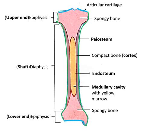
Table of Contents
The Ends of a Long Bone Are Called the Epiphyses: A Deep Dive into Bone Anatomy and Growth
The ends of a long bone are called the epiphyses (singular: epiphysis). Understanding the epiphyses is crucial to comprehending bone growth, development, and various skeletal conditions. This comprehensive article will delve into the intricate anatomy of the epiphyses, their role in bone growth, common injuries and disorders affecting this region, and the diagnostic methods employed to assess their health.
What are Epiphyses?
Epiphyses are the secondary ossification centers located at the ends of long bones. Unlike the diaphysis (the shaft of the long bone), which ossifies earlier, the epiphyses begin ossification later during development. This process is crucial for longitudinal bone growth. The epiphysis is primarily composed of spongy bone, also known as cancellous bone, which is a porous, lightweight type of bone tissue. This spongy structure provides ample space for bone marrow, which plays a critical role in hematopoiesis (blood cell production). The epiphysis is covered by a thin layer of hyaline cartilage, called articular cartilage, which allows for smooth, low-friction movement at the joints.
Distinguishing the Epiphysis from Other Bone Structures:
It’s important to distinguish the epiphysis from other related structures:
- Metaphysis: This is the transitional region between the epiphysis and the diaphysis. It contains the growth plate, also known as the physis, a crucial area for longitudinal bone growth.
- Diaphysis: The long, cylindrical shaft of the bone. It's primarily composed of compact bone, a dense and strong type of bone tissue.
- Articular Cartilage: The smooth, white tissue covering the epiphysis at the joint surface, enabling frictionless joint movement.
The Role of Epiphyses in Bone Growth:
The epiphyseal growth plate, located within the metaphysis, is the site of longitudinal bone growth. This plate consists of specialized cartilage cells that continuously divide and differentiate, leading to an increase in bone length. This process, called endochondral ossification, involves the gradual replacement of cartilage with bone tissue. The growth plate remains active until puberty, when it eventually closes, marking the end of longitudinal bone growth. The closure of the epiphyseal plate is a complex process influenced by various hormonal factors, primarily growth hormone and sex hormones.
Understanding Endochondral Ossification: A Step-by-Step Process
- Chondrocyte Proliferation: Cartilage cells (chondrocytes) in the growth plate actively divide and proliferate.
- Cartilage Matrix Deposition: New cartilage matrix is laid down, pushing the epiphysis further away from the diaphysis.
- Hypertrophy and Calcification: Chondrocytes mature, enlarge (hypertrophy), and the surrounding cartilage matrix calcifies.
- Osteoblast Invasion: Blood vessels invade the calcified cartilage, bringing osteoblasts, which are bone-forming cells.
- Bone Formation: Osteoblasts deposit new bone tissue, replacing the calcified cartilage.
This continuous cycle of cartilage production, calcification, and bone replacement leads to the lengthening of the bone. Once the growth plate closes, further longitudinal growth is impossible. The remnants of the growth plate become visible as the epiphyseal line, a radiologically visible line indicating the fusion of the epiphysis and diaphysis.
Common Injuries and Disorders Affecting the Epiphyses:
Several injuries and disorders can affect the epiphyses, often with significant consequences for bone growth and development:
1. Epiphyseal Fractures:
These fractures occur within the growth plate and can have devastating consequences, potentially leading to growth disturbances. The Salter-Harris classification system is widely used to categorize epiphyseal fractures based on the involvement of the growth plate. Type I fractures involve the growth plate only, while Type V fractures involve compression of the growth plate. The severity of the injury and the potential for growth disturbances depend on the fracture type and the effectiveness of treatment.
2. Slipped Capital Femoral Epiphysis (SCFE):
This condition involves the displacement of the femoral head (the ball of the hip joint) from the neck of the femur. It typically affects adolescents during puberty and can severely impair hip function and growth. Early diagnosis and intervention are crucial to prevent long-term complications.
3. Osteochondritis Dissecans (OCD):
OCD involves the separation of a segment of articular cartilage and underlying bone from the epiphysis. This condition commonly affects the knee and elbow joints and can cause pain, swelling, and joint instability. Treatment varies depending on the severity and may involve surgery in some cases.
4. Legg-Calvé-Perthes Disease:
This condition affects the blood supply to the femoral head, leading to avascular necrosis (death of bone tissue). It primarily affects young children and can result in significant hip deformity if left untreated.
5. Achondroplasia:
This is a genetic disorder affecting bone growth, resulting in dwarfism. Individuals with achondroplasia have shortened limbs due to abnormalities in endochondral ossification at the epiphyseal growth plates.
Diagnostic Methods for Assessing Epiphyseal Health:
Several imaging techniques are used to assess the health and integrity of the epiphyses:
- X-rays: X-rays provide excellent visualization of bone structures, including the epiphyses, growth plates, and epiphyseal lines. They are essential in diagnosing fractures, assessing growth plate injuries, and evaluating the extent of bone abnormalities.
- MRI (Magnetic Resonance Imaging): MRI provides detailed images of soft tissues, including cartilage, making it invaluable in diagnosing conditions such as OCD and assessing the integrity of the articular cartilage.
- CT (Computed Tomography): CT scans offer high-resolution images of bone and can be useful in evaluating complex fractures or bone abnormalities involving the epiphyses.
- Bone Scans: Bone scans can detect areas of increased metabolic activity, which may indicate bone inflammation or injury.
Conclusion:
The epiphyses are vital components of long bones, playing a crucial role in bone growth and development. Understanding their anatomy, function, and susceptibility to injury and disease is essential for healthcare professionals involved in the diagnosis and management of musculoskeletal disorders. Early detection and appropriate treatment are crucial in minimizing the long-term consequences of epiphyseal injuries and diseases, ensuring optimal skeletal health and function. Further research continues to explore the complex processes governing epiphyseal growth and development, paving the way for improved diagnostic tools and therapeutic interventions. This deeper understanding of the epiphyses allows for better preventative measures and improved outcomes for patients affected by related conditions. The continuous advancements in imaging technologies and our understanding of bone biology continue to improve the management and treatment of epiphyseal-related pathologies.
Latest Posts
Latest Posts
-
Current As A Function Of Time
Mar 18, 2025
-
Ode To Billy Joe Lyrics Meaning
Mar 18, 2025
-
Where Does The Light Independent Reaction Take Place
Mar 18, 2025
-
S P D F Blocks On The Periodic Table
Mar 18, 2025
-
Delta G Of A Carbonyl Reduction
Mar 18, 2025
Related Post
Thank you for visiting our website which covers about The Ends Of A Long Bone Are Called The . We hope the information provided has been useful to you. Feel free to contact us if you have any questions or need further assistance. See you next time and don't miss to bookmark.
