The Mitotic Spindles Arise From Which Cell Structure
Muz Play
Mar 31, 2025 · 6 min read

Table of Contents
The Mitotic Spindles Arise From Which Cell Structure? A Deep Dive into Centrosomes and Microtubule Organization
The precise orchestration of chromosome segregation during cell division is a fundamental process for life. Central to this intricate dance is the mitotic spindle, a dynamic, bipolar structure responsible for separating duplicated chromosomes into daughter cells. But the question remains: from which cell structure do these crucial mitotic spindles arise? The answer lies within the centrosome, a complex organelle acting as the primary microtubule-organizing center (MTOC) in many animal cells. This article will delve deep into the centrosome's structure, function, and its crucial role in mitotic spindle formation, exploring the intricate interplay of proteins and microtubules that make this process possible. We'll also touch upon variations in spindle formation in organisms lacking centrosomes, showcasing the adaptability of this essential cellular mechanism.
Understanding the Centrosome: The Microtubule Organizing Center
The centrosome, often described as the "cell's microtubule-organizing center," is a small, electron-dense structure typically located near the nucleus. It's far from a static entity; rather, it's a highly dynamic organelle whose composition and activity change throughout the cell cycle. Critically, its core function is to nucleate and organize microtubules, the protein polymers forming the structural backbone of the mitotic spindle.
Centrosome Structure: A Pair of Centrioles Embedded in Pericentriolar Material (PCM)
A key feature distinguishing the centrosome is its structural organization. At its heart lie two cylindrical structures called centrioles, arranged perpendicularly to each other. These centrioles, themselves composed of nine triplet microtubules arranged in a cartwheel pattern, are embedded within a pericentriolar material (PCM). This PCM is a proteinaceous matrix containing numerous proteins crucial for microtubule nucleation, anchoring, and regulation. The PCM's composition is incredibly complex, with hundreds of proteins identified, each contributing to the centrosome's multifaceted functions. Key proteins include:
- γ-tubulin ring complex (γ-TuRC): This crucial complex serves as the template for microtubule nucleation, initiating the polymerization of α- and β-tubulin dimers into microtubules. Its presence in the PCM is essential for the centrosome's role as the MTOC.
- Pericentrin and Centrosomin: These proteins act as scaffolding proteins, contributing to the structural integrity of the PCM and regulating the recruitment of other proteins.
- Ninein: This protein plays a role in linking the centrioles to the PCM, ensuring the structural stability of the centrosome.
- CDK5RAP2: This kinase regulator is essential for centrosome maturation and duplication.
The dynamic interplay of these proteins ensures the proper assembly, positioning, and function of the centrosome. Any disruption in this intricate network can have severe consequences, leading to errors in chromosome segregation and potentially causing aneuploidy – an abnormal number of chromosomes in cells.
Centrosome Duplication: A Precise Process Ensuring Accurate Chromosome Segregation
The centrosome, like other cellular organelles, undergoes duplication during the cell cycle. This process, tightly coupled with the cell cycle, ensures that each daughter cell receives a centrosome, essential for organizing the microtubule arrays required for subsequent cell divisions. Centrosome duplication begins during the S phase of the cell cycle, alongside DNA replication. The process is carefully regulated to prevent numerical errors that could lead to genomic instability. Key steps include:
- Centriole duplication: Each centriole acts as a template for the formation of a new, daughter centriole. This process involves the recruitment and assembly of specific proteins at the proximal end of the mother centriole, gradually forming a new centriole.
- PCM recruitment and expansion: As the centrioles duplicate, the PCM surrounding them also expands and matures. This expansion requires the recruitment and assembly of numerous proteins, leading to the formation of two fully functional centrosomes.
- Centrosome separation: During mitosis, the duplicated centrosomes separate, migrating to opposite poles of the cell. This migration is crucial for establishing the bipolar spindle, necessary for accurate chromosome segregation. The process involves motor proteins, such as dynein and kinesin, that actively move the centrosomes along microtubules.
The precise regulation of centrosome duplication is crucial for maintaining genomic stability. Errors in this process can lead to abnormal centrosome numbers, resulting in multipolar spindles, which can lead to chromosome mis-segregation and aneuploidy, potentially contributing to cancer development.
The Centrosome's Role in Mitotic Spindle Assembly
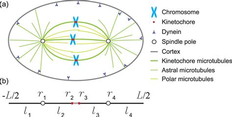
The centrosome plays a central role in mitotic spindle assembly, acting as the primary MTOC. The process is complex, involving numerous proteins and intricate interactions between microtubules and other cellular components. Here’s a breakdown of the key events:
Microtubule Nucleation and Elongation
The centrosome, rich in γ-TuRC complexes, serves as the primary site for microtubule nucleation. The γ-TuRC acts as a template, initiating the polymerization of α- and β-tubulin dimers into microtubules that radiate outward from the centrosome. These microtubules, initially short and unstable, elongate and become stabilized through interactions with various microtubule-associated proteins (MAPs).
Centrosome Separation and Bipolar Spindle Formation
As the cell progresses into mitosis, the duplicated centrosomes separate and migrate to opposite poles of the cell, driven by motor proteins and microtubule dynamics. This separation establishes the two poles of the mitotic spindle, forming a bipolar structure.
Chromosome Capture and Alignment
Microtubules emanating from the centrosomes then capture chromosomes via kinetochores, specialized protein structures located at the centromeres of chromosomes. This attachment is essential for proper chromosome segregation. The spindle microtubules attach to kinetochores and exert forces that align chromosomes along the metaphase plate, ensuring that each chromosome is properly oriented for segregation.
Chromosome Segregation and Cytokinesis
Once chromosomes are correctly aligned, the spindle apparatus undergoes a series of dynamic changes, pulling sister chromatids apart towards opposite poles of the cell. This separation is followed by cytokinesis, the physical division of the cell into two daughter cells, each receiving a complete set of chromosomes and a centrosome.
Mitotic Spindle Formation in Acentrosomal Cells: Alternative Mechanisms
While the centrosome is the primary MTOC in many animal cells, it's important to note that not all eukaryotic cells rely on centrosomes for mitotic spindle formation. Many plants, fungi, and some animal cells assemble spindles in a centrosome-independent manner. In these acentrosomal cells, microtubule nucleation occurs at multiple sites within the cytoplasm, leading to the formation of a bipolar spindle through different mechanisms, including:
- Chromosomes as MTOCs: In some acentrosomal cells, the chromosomes themselves can nucleate microtubules, contributing to spindle formation.
- Ran GTPase-mediated nucleation: RanGTP, a small GTPase, plays a crucial role in promoting microtubule nucleation in the cytoplasm, independent of centrosomes.
- Microtubule self-organization: Microtubules can self-organize into bipolar spindles through interactions between microtubules and motor proteins.
Conclusion: The Centrosome, a Master Regulator of Cell Division
The mitotic spindle, a marvel of cellular organization, arises primarily from the centrosome, a dynamic organelle acting as the cell's MTOC. The centrosome's precise duplication, its role in microtubule nucleation, and its contribution to spindle organization are crucial for accurate chromosome segregation. While the centrosome is central in many organisms, acentrosomal spindle formation demonstrates the inherent adaptability of this vital cellular process. Further research into the intricacies of centrosome biology and spindle formation promises to yield invaluable insights into cell division, cell cycle regulation, and the development of strategies to combat diseases arising from chromosome instability, highlighting the crucial role of this often-overlooked cellular structure.
Latest Posts
Latest Posts
-
Induced Fit Model Vs Lock And Key
Apr 02, 2025
-
Colligative Properties Freezing Point Depression Lab
Apr 02, 2025
-
Matrix As A Product Of Elementary Matrices
Apr 02, 2025
-
Is Ethnic Studies A Social Science
Apr 02, 2025
-
Name The Structural And Functional Unit Of All Living Things
Apr 02, 2025
Related Post
Thank you for visiting our website which covers about The Mitotic Spindles Arise From Which Cell Structure . We hope the information provided has been useful to you. Feel free to contact us if you have any questions or need further assistance. See you next time and don't miss to bookmark.
