The Plasma Membrane Of A Muscle Fiber Is Called The
Muz Play
Mar 23, 2025 · 6 min read
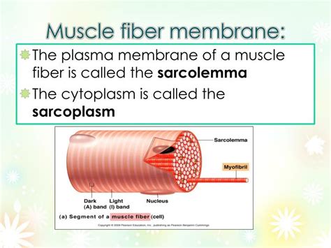
Table of Contents
The Plasma Membrane of a Muscle Fiber is Called the Sarcolemma: A Deep Dive into Muscle Cell Structure and Function
The intricate workings of the human body are a testament to the elegance of biological design. At the heart of movement and locomotion lies the muscle fiber, a highly specialized cell responsible for generating force. Understanding the structure and function of this cell is crucial to comprehending how we move, and a key component of this understanding lies in its plasma membrane. But what is the plasma membrane of a muscle fiber actually called? The answer is the sarcolemma. This article delves deep into the sarcolemma, exploring its structure, composition, and crucial role in muscle fiber function, encompassing the excitation-contraction coupling process.
What is the Sarcolemma?
The sarcolemma, derived from the Greek words "sarx" (flesh) and "lemma" (sheath), is the specialized plasma membrane that encloses a muscle fiber (myofiber). It's much more than just a simple boundary; it's a dynamic structure intricately involved in the complex processes that lead to muscle contraction. Unlike the plasma membranes of other cells, the sarcolemma exhibits unique structural features optimized for rapid and efficient signal transmission, crucial for the coordinated action of muscle fibers.
The Sarcolemma's Unique Structure:
The sarcolemma isn't a uniform membrane; it comprises several distinct components working in concert:
-
Plasma Membrane: The fundamental lipid bilayer, composed of phospholipids, cholesterol, and proteins, provides the basic structural framework. This layer acts as a selective barrier, regulating the passage of ions and molecules into and out of the muscle fiber.
-
Basement Membrane: A thin layer of extracellular matrix that lies beneath the plasma membrane, providing structural support and anchoring the muscle fiber to surrounding connective tissue. It also plays a role in regulating the muscle fiber's environment and interactions with other cells.
-
Transverse Tubules (T-tubules): These are invaginations of the sarcolemma that extend deep into the muscle fiber, forming a network that ensures rapid and uniform spread of the action potential throughout the myofiber. This is vital for synchronous contraction of the myofibrils within the muscle fiber.
-
Specialized Proteins: The sarcolemma is studded with a variety of membrane proteins, including ion channels, transporters, and receptors. These proteins are critical for the transmission of electrical signals, the regulation of calcium ion concentration, and the maintenance of membrane potential. Specifically, voltage-gated sodium and potassium channels are crucial for the propagation of the action potential, while calcium channels and transporters are involved in regulating intracellular calcium levels, a key factor in muscle contraction.
The Sarcolemma's Role in Excitation-Contraction Coupling
The sarcolemma plays a pivotal role in excitation-contraction coupling (ECC), the process by which an electrical signal (action potential) triggers muscle contraction. This intricate sequence of events involves a precise interplay between the sarcolemma and the internal structures of the muscle fiber:
-
Action Potential Arrival: A nerve impulse reaches the neuromuscular junction, triggering the release of acetylcholine. This neurotransmitter binds to receptors on the sarcolemma, initiating depolarization—a change in the membrane potential that makes the inside of the cell more positive.
-
Action Potential Propagation: The depolarization wave travels along the sarcolemma and into the T-tubules, rapidly spreading the signal throughout the entire muscle fiber. This rapid spread ensures that all myofibrils within the fiber contract simultaneously.
-
Calcium Release: The depolarization wave reaching the T-tubules triggers the release of calcium ions (Ca²⁺) from the sarcoplasmic reticulum (SR), a specialized intracellular calcium store. This calcium release is mediated by voltage-sensitive proteins located at the junction between the T-tubules and the SR, a structure called the triad.
-
Muscle Contraction: The released calcium ions bind to troponin, a protein complex on the actin filaments. This binding initiates a conformational change in troponin, exposing the myosin-binding sites on actin. Myosin heads then bind to actin, initiating the cross-bridge cycle and ultimately leading to muscle contraction.
-
Relaxation: Once the action potential ceases, calcium ions are actively pumped back into the SR via the sarco/endoplasmic reticulum Ca²⁺-ATPase (SERCA) pump. The removal of calcium ions from the cytoplasm causes the troponin to return to its resting conformation, blocking the myosin-binding sites on actin, leading to muscle relaxation.
The sarcolemma's role in this process is absolutely vital. Without its ability to conduct the action potential rapidly and efficiently, muscle contraction would be uncoordinated and weak. The intricate architecture of the T-tubules, in particular, is crucial for ensuring uniform signal transmission throughout the muscle fiber.
Sarcolemma and Muscle Fiber Types
The structure and function of the sarcolemma can vary slightly depending on the type of muscle fiber. Skeletal muscle fibers are broadly classified into different types based on their contractile properties, and these variations are reflected in the characteristics of their sarcolemma:
-
Type I (Slow-twitch) Fibers: These fibers are specialized for endurance activities. Their sarcolemma may contain a higher density of certain ion channels and transporters that support their sustained, low-intensity contractions.
-
Type II (Fast-twitch) Fibers: These fibers are designed for rapid, powerful contractions. Their sarcolemma might exhibit characteristics that facilitate faster action potential propagation and calcium handling, enhancing their speed and power output. Type II fibers are further subdivided into Type IIa (fast-oxidative-glycolytic) and Type IIx (fast-glycolytic) fibers, each with slightly different sarcolemma properties.
The specific composition of ion channels, transporters, and receptors within the sarcolemma helps tailor the muscle fiber's response to different stimuli, allowing for the diverse range of movements the human body is capable of.
Diseases Affecting the Sarcolemma
Several diseases can impair the structure and function of the sarcolemma, leading to various muscle disorders. These conditions can impact the sarcolemma's ability to conduct electrical signals, regulate ion transport, or maintain its structural integrity. Some notable examples include:
-
Muscular Dystrophies: A group of genetic disorders characterized by progressive muscle weakness and degeneration. Many forms of muscular dystrophy involve mutations in genes encoding proteins crucial for maintaining the structural integrity of the sarcolemma, leading to membrane damage and muscle fiber death.
-
Myasthenia Gravis: An autoimmune disorder in which antibodies attack the acetylcholine receptors on the sarcolemma at the neuromuscular junction. This reduces the effectiveness of neuromuscular transmission, leading to muscle weakness and fatigue.
-
Lambert-Eaton Myasthenic Syndrome (LEMS): Another autoimmune disorder targeting voltage-gated calcium channels on the presynaptic nerve terminals. This affects the release of acetylcholine, leading to muscle weakness.
-
Periodic Paralysis: A group of disorders characterized by episodes of muscle weakness or paralysis, often triggered by changes in potassium levels. These conditions may involve abnormalities in ion channels within the sarcolemma.
Understanding the role of the sarcolemma in these diseases is crucial for developing effective diagnostic and therapeutic strategies.
Conclusion: The Sarcolemma – A Dynamic Player in Muscle Physiology
The sarcolemma, the plasma membrane of a muscle fiber, is far more than a simple cellular boundary. Its intricate structure, including the T-tubules and associated proteins, plays a critical role in the excitation-contraction coupling process. This dynamic structure is responsible for the rapid and efficient transmission of electrical signals throughout the muscle fiber, triggering the release of calcium and ultimately leading to muscle contraction. Moreover, variations in sarcolemma composition contribute to the diverse functional properties of different muscle fiber types. The study of the sarcolemma is essential for understanding normal muscle function and the pathogenesis of various muscle disorders. Further research into its intricate workings will undoubtedly continue to reveal new insights into muscle physiology and potential therapeutic targets for muscle diseases. The sarcolemma's importance in muscle function cannot be overstated; it is the gateway to the power of movement.
Latest Posts
Latest Posts
-
Table Of Elements With Protons Neutrons And Electrons
Mar 25, 2025
-
The Level With The Most Energy Is The Level
Mar 25, 2025
-
Fcc Unit Cell Number Of Atoms
Mar 25, 2025
-
How Is Temperature Related To Kinetic Energy
Mar 25, 2025
-
Which Element Has The Largest Ionization Energy
Mar 25, 2025
Related Post
Thank you for visiting our website which covers about The Plasma Membrane Of A Muscle Fiber Is Called The . We hope the information provided has been useful to you. Feel free to contact us if you have any questions or need further assistance. See you next time and don't miss to bookmark.
