Vascular Anatomy Of The Lower Extremity
Muz Play
Mar 26, 2025 · 6 min read
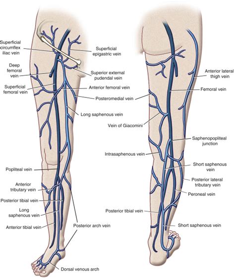
Table of Contents
- Vascular Anatomy Of The Lower Extremity
- Table of Contents
- Vascular Anatomy of the Lower Extremity: A Comprehensive Guide
- Arterial Supply of the Lower Extremity
- Femoral Artery and its Branches
- Popliteal Artery
- Anterior and Posterior Tibial Arteries
- Venous Drainage of the Lower Extremity
- Superficial Veins
- Deep Veins
- Venous Valves
- Lymphatic Drainage of the Lower Extremity
- Clinical Significance
- Diagnostic Imaging Techniques
- Conclusion
- Latest Posts
- Latest Posts
- Related Post
Vascular Anatomy of the Lower Extremity: A Comprehensive Guide
The lower extremity's vascular system is a complex network responsible for delivering oxygenated blood to the muscles, tissues, and organs of the legs and feet, and returning deoxygenated blood to the heart. Understanding its intricate anatomy is crucial for clinicians, researchers, and students alike. This comprehensive guide will delve into the arterial, venous, and lymphatic systems of the lower extremity, providing a detailed overview of their structure, function, and clinical significance.
Arterial Supply of the Lower Extremity
The arterial supply originates from the external iliac artery, which continues as the femoral artery upon entering the thigh. This is the major artery of the lower extremity. Let's explore its branches and their distribution:
Femoral Artery and its Branches
The femoral artery, located in the femoral triangle, gives rise to several important branches:
- Superficial epigastric artery: Supplies the superficial tissues of the lower abdomen.
- Superficial circumflex iliac artery: Supplies the superficial tissues of the iliac region.
- Superficial external pudendal artery: Supplies the external genitalia.
- Deep external pudendal artery: Also contributes to the blood supply of the external genitalia.
- Profunda femoris artery (deep femoral artery): A crucial branch supplying the deep muscles of the thigh. It further branches into the medial and lateral circumflex femoral arteries, perfusing the hip joint and surrounding muscles. The profunda femoris artery also gives off perforating arteries that supply the posterior compartment of the thigh.
Popliteal Artery
As the femoral artery passes through the adductor hiatus, it enters the popliteal fossa and becomes the popliteal artery. This artery is located deep within the popliteal fossa, surrounded by muscles and nerves. Its branches supply the knee joint and surrounding muscles.
Anterior and Posterior Tibial Arteries
The popliteal artery divides into the anterior tibial artery and the tibial trunk. The tibial trunk then bifurcates into the posterior tibial artery and the fibular (peroneal) artery.
-
Anterior tibial artery: This artery runs anterior to the interosseous membrane, supplying the anterior compartment of the leg. It eventually becomes the dorsalis pedis artery at the ankle. The dorsalis pedis artery is a clinically important landmark for assessing peripheral circulation. It gives off branches to the dorsum of the foot and toes.
-
Posterior tibial artery: Situated posterior to the interosseous membrane, this artery supplies the posterior compartment of the leg and gives off the medial plantar artery and lateral plantar artery in the foot, forming the plantar arch. The plantar arch provides the blood supply to the plantar aspect of the foot and toes.
-
Fibular (peroneal) artery: Running along the fibula, this artery supplies the lateral compartment of the leg and contributes to the blood supply of the foot.
Venous Drainage of the Lower Extremity
The venous system of the lower extremity is crucial for returning deoxygenated blood to the heart. It's characterized by a superficial and deep system:
Superficial Veins
The superficial veins lie just beneath the skin and are responsible for collecting blood from the skin and subcutaneous tissues. The major superficial veins include:
- Great saphenous vein: The longest vein in the body, it begins on the medial side of the foot, ascends along the medial aspect of the leg and thigh, and drains into the femoral vein.
- Small saphenous vein: Originates on the lateral side of the foot, ascends along the posterior aspect of the leg, and drains into the popliteal vein.
Deep Veins
The deep veins run alongside the arteries, sharing similar names and anatomical locations. They collect blood from the deep muscles and tissues. The major deep veins include:
- Femoral vein: Receives blood from the deep veins of the thigh and the great saphenous vein.
- Popliteal vein: Formed by the union of the anterior and posterior tibial veins.
- Anterior and posterior tibial veins: Drain the corresponding compartments of the leg.
- Fibular (peroneal) veins: Drain the lateral compartment of the leg.
- External iliac vein: Continues from the femoral vein and ultimately contributes to the inferior vena cava.
Venous Valves
Both superficial and deep veins possess valves that prevent backflow of blood, ensuring unidirectional flow towards the heart. The impairment of these valves can lead to venous insufficiency and varicose veins.
Lymphatic Drainage of the Lower Extremity
The lymphatic system plays a vital role in immune defense and fluid balance. Lymphatic vessels in the lower extremity collect lymph fluid from the tissues and transport it to lymph nodes, where immune cells filter and process the fluid. The lymph nodes are strategically located throughout the lower extremity, filtering lymph before it flows to larger lymphatic trunks.
Clinical Significance
Understanding the vascular anatomy of the lower extremity is vital for diagnosing and managing various conditions, including:
- Peripheral artery disease (PAD): Characterized by atherosclerosis in the arteries of the lower extremities, leading to reduced blood flow. Symptoms include intermittent claudication, rest pain, and non-healing ulcers.
- Deep vein thrombosis (DVT): Formation of blood clots in the deep veins, potentially leading to pulmonary embolism.
- Venous insufficiency: Impaired venous return, resulting in edema, varicose veins, and skin changes.
- Lymphedema: Accumulation of lymphatic fluid due to lymphatic obstruction.
- Diabetic foot ulcers: Chronic wounds that often occur in patients with diabetes due to peripheral neuropathy and impaired circulation.
- Trauma and injuries: Understanding the vascular anatomy helps in assessing and managing injuries to blood vessels and nerves.
Diagnostic Imaging Techniques
Several imaging techniques are used to visualize the vascular system of the lower extremity:
- Doppler ultrasound: A non-invasive method used to assess blood flow in arteries and veins.
- Angiography: Invasive procedure involving injecting contrast dye into the arteries to visualize them using X-ray.
- Magnetic resonance angiography (MRA): Non-invasive imaging technique using magnetic resonance to visualize blood vessels.
- Computed tomography angiography (CTA): Non-invasive imaging technique using computed tomography to visualize blood vessels.
Conclusion
The vascular anatomy of the lower extremity is a complex yet fascinating system essential for maintaining the health and function of the legs and feet. A thorough understanding of its arterial, venous, and lymphatic components is crucial for diagnosing and treating a wide range of vascular disorders. Clinicians, researchers, and students alike should continue to explore and refine their knowledge of this intricate network to improve patient care and outcomes. Further research into the intricate interplay between the various components of the lower extremity vascular system will undoubtedly lead to advancements in diagnosis and treatment of vascular pathologies. The ongoing development of imaging techniques promises to further enhance our ability to visualize and understand this crucial anatomical region. The continuous exploration and integration of this knowledge into clinical practice are paramount for optimizing patient care and overall health. The complex interplay between the arterial, venous, and lymphatic systems highlights the need for a holistic approach to the understanding and management of lower extremity vascular health.
Latest Posts
Latest Posts
-
How Do C4 Plants Minimize Photorespiration
Mar 27, 2025
-
How To Divide Fractions With Exponents
Mar 27, 2025
-
How Many Valence Electrons In Be
Mar 27, 2025
-
Primer Design For Site Directed Mutagenesis
Mar 27, 2025
-
Is Carbon Dioxide A Pure Substance Or A Mixture
Mar 27, 2025
Related Post
Thank you for visiting our website which covers about Vascular Anatomy Of The Lower Extremity . We hope the information provided has been useful to you. Feel free to contact us if you have any questions or need further assistance. See you next time and don't miss to bookmark.
