What Are The Two Divisions Of The Skeletal System
Muz Play
Mar 24, 2025 · 7 min read
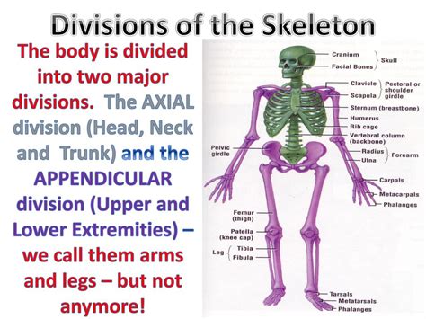
Table of Contents
The Two Divisions of the Skeletal System: A Deep Dive into Axial and Appendicular Structures
The human skeletal system, a marvel of biological engineering, provides the structural framework for our bodies. Far from being a static collection of bones, it's a dynamic and interconnected system crucial for movement, protection of vital organs, blood cell production, and mineral storage. Understanding its structure is key to appreciating its complex functions. This comprehensive guide delves into the two major divisions of the skeletal system: the axial and appendicular skeletons, exploring their individual components, functions, and interrelationships.
The Axial Skeleton: The Body's Central Support Structure
The axial skeleton forms the central axis of the body. Think of it as the core, providing the foundational support for the appendicular skeleton and protecting vital organs. It's composed of 80 bones, roughly 40% of the total bones in the adult human body. These bones can be categorized into three main regions: the skull, the vertebral column, and the thoracic cage.
1. The Skull: Protecting the Brain and Sensory Organs
The skull, arguably the most recognizable part of the axial skeleton, is a complex structure comprised of 22 bones. These bones are expertly fused together (except for the mandible, or lower jaw) to form a protective enclosure for the brain and crucial sensory organs. We can further subdivide the skull into two main parts: the cranium and the facial bones.
-
The Cranium (Neurocranium): This bony vault protects the brain. It comprises eight major bones: the frontal bone (forehead), two parietal bones (forming the sides and top of the skull), two temporal bones (housing the inner ear and parts of the jaw joint), the occipital bone (forming the back of the skull and containing the foramen magnum, the large opening where the spinal cord exits the brain), the sphenoid bone (a complex, bat-shaped bone that forms part of the base of the skull and eye sockets), and the ethmoid bone (contributing to the nasal cavity and eye sockets). The intricate sutures connecting these bones are critical for flexibility during birth and for overall cranial strength.
-
The Facial Bones (Viscerocranium): These 14 bones provide structure to the face, supporting the eyes, nose, and mouth. Prominent facial bones include the mandible (lower jawbone, the only freely movable bone in the skull), the maxillae (upper jawbones, forming the upper part of the mouth and nasal cavity), the zygomatic bones (cheekbones), the nasal bones (forming the bridge of the nose), and the lacrimal bones (small bones contributing to the medial wall of each eye socket). These bones also play a vital role in speech, chewing, and facial expression.
2. The Vertebral Column: Flexibility and Support
The vertebral column, also known as the spine or backbone, is a flexible, yet strong, column of 33 vertebrae. These bones are stacked upon one another, forming a vertical axis that supports the head, neck, and torso, whilst protecting the delicate spinal cord. The vertebrae are grouped into five regions:
-
Cervical Vertebrae (C1-C7): Seven vertebrae in the neck, characterized by their small size and specific features that allow for significant range of motion. The first two vertebrae, the atlas (C1) and axis (C2), are uniquely shaped to allow for head rotation and nodding.
-
Thoracic Vertebrae (T1-T12): Twelve vertebrae in the chest region, each articulating with ribs to form the thoracic cage. These vertebrae are larger and more robust than the cervical vertebrae, providing stability for the rib cage.
-
Lumbar Vertebrae (L1-L5): Five vertebrae in the lower back, the largest and strongest vertebrae in the spine, designed to bear the weight of the upper body.
-
Sacrum: Five fused vertebrae forming a triangular bone at the base of the spine, connecting the vertebral column to the pelvis.
-
Coccyx: Four fused vertebrae forming the tailbone, a vestigial structure representing the remnant of a tail.
Intervertebral discs, made of cartilage, sit between the vertebrae, acting as shock absorbers and facilitating movement. The curvature of the spine is vital for posture, shock absorption, and overall skeletal integrity.
3. The Thoracic Cage: Protecting Vital Organs
The thoracic cage, also called the rib cage, is a bony structure composed of 12 pairs of ribs, the sternum (breastbone), and the thoracic vertebrae. It encloses and protects vital organs such as the heart and lungs.
-
Ribs: Seven pairs of true ribs attach directly to the sternum via costal cartilage. Three pairs of false ribs attach indirectly to the sternum through cartilage connected to the seventh rib. Two pairs of floating ribs do not attach to the sternum at all.
-
Sternum: A flat, elongated bone located in the anterior chest wall. It's divided into three parts: the manubrium, the body, and the xiphoid process.
The Appendicular Skeleton: Movement and Manipulation
The appendicular skeleton comprises the bones of the limbs (appendages) and their supporting structures, enabling locomotion, manipulation of objects, and a wide range of movements. It consists of 126 bones, approximately 60% of the total bones in the adult human body. It is subdivided into the pectoral girdle, upper limbs, pelvic girdle, and lower limbs.
1. The Pectoral Girdle: Connecting the Upper Limbs to the Axial Skeleton
The pectoral girdle, or shoulder girdle, connects each upper limb to the axial skeleton. It's formed by two clavicles (collarbones) and two scapulae (shoulder blades). These bones allow for a wide range of motion in the upper limbs. The relatively loose connection of the pectoral girdle to the axial skeleton contributes significantly to the upper limb’s flexibility.
2. The Upper Limbs: Precision and Dexterity
Each upper limb consists of 30 bones, organized into three main sections:
-
Humerus: The long bone of the upper arm.
-
Radius and Ulna: Two bones in the forearm, the radius on the thumb side, and the ulna on the pinky finger side. Their articulation allows for pronation and supination (rotating the forearm).
-
Carpals, Metacarpals, and Phalanges: The wrist (carpals), palm (metacarpals), and fingers (phalanges) respectively. The hand's intricate structure allows for fine motor control and dexterity.
3. The Pelvic Girdle: Supporting the Lower Limbs and Protecting Internal Organs
The pelvic girdle, or hip girdle, is a strong, stable structure formed by two hip bones (ossa coxae). Each hip bone is formed by the fusion of three bones: the ilium, ischium, and pubis. The pelvic girdle connects the lower limbs to the axial skeleton, providing support for the weight of the upper body and protecting internal organs such as the bladder, rectum, and reproductive organs. The pelvic girdle is significantly stronger and less mobile than the pectoral girdle, reflecting its role in weight-bearing and stability.
4. The Lower Limbs: Locomotion and Stability
Each lower limb comprises 30 bones, arranged into three main sections:
-
Femur: The thigh bone, the longest and strongest bone in the body.
-
Patella: The kneecap, a sesamoid bone (bone embedded in a tendon) that protects the knee joint.
-
Tibia and Fibula: Two bones in the lower leg, the tibia (shin bone) bearing most of the body's weight, and the fibula providing lateral stability.
-
Tarsals, Metatarsals, and Phalanges: The ankle bones (tarsals), the bones of the foot (metatarsals), and the bones of the toes (phalanges). The foot's structure is adapted for weight-bearing, shock absorption, and locomotion.
Interrelation Between Axial and Appendicular Skeletons
The axial and appendicular skeletons are not isolated entities; they work together seamlessly. The axial skeleton provides the central framework, while the appendicular skeleton extends this framework, enabling movement and manipulation. The connection points between these two divisions are critical for overall body function. For example:
-
Shoulder and Hip Joints: The pectoral and pelvic girdles connect the appendicular skeleton to the axial skeleton, allowing for the movement of the limbs.
-
Vertebral Column: The vertebral column supports the weight of the head and upper body, while also transmitting forces from the lower limbs during movement.
-
Rib Cage and Pelvic Girdle: These structures protect vital organs and provide attachment points for muscles involved in respiration and locomotion.
Clinical Significance and Conclusion
Understanding the structure and function of the axial and appendicular skeletons is essential in various medical fields. Conditions affecting the skeletal system, such as fractures, osteoporosis, scoliosis, and arthritis, can significantly impact an individual's mobility, quality of life, and overall health. Accurate diagnosis and treatment rely heavily on a comprehensive understanding of the skeletal system's anatomy.
In conclusion, the human skeletal system, divided into the axial and appendicular skeletons, is a remarkable structure. The axial skeleton provides the body's central support and protects vital organs, while the appendicular skeleton facilitates movement and manipulation. The intricate interplay between these two divisions allows us to perform the complex movements necessary for daily life. A thorough understanding of their individual components and their interconnectedness is crucial for appreciating the complexity and functionality of this remarkable system.
Latest Posts
Latest Posts
-
Directional Selection Disruptive Selection Stabilizing Selection
Mar 26, 2025
-
Is Blood Clotting A Positive Or Negative Feedback
Mar 26, 2025
-
How To Calculate The Expected Frequency
Mar 26, 2025
-
Positive And Increasing Rate Of Change
Mar 26, 2025
-
Anything That Has Mass And Volume Is Called
Mar 26, 2025
Related Post
Thank you for visiting our website which covers about What Are The Two Divisions Of The Skeletal System . We hope the information provided has been useful to you. Feel free to contact us if you have any questions or need further assistance. See you next time and don't miss to bookmark.
