What Are The Two Divisions Of The Skeleton
Muz Play
Mar 23, 2025 · 8 min read
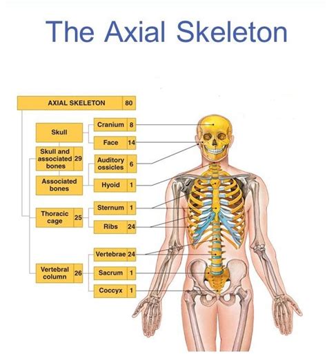
Table of Contents
- What Are The Two Divisions Of The Skeleton
- Table of Contents
- What Are the Two Divisions of the Skeleton? A Comprehensive Guide
- The Axial Skeleton: The Body's Central Support Structure
- 1. The Skull: Protecting the Brain and Sensory Organs
- 2. The Vertebral Column: Flexibility and Protection of the Spinal Cord
- 3. The Thoracic Cage: Protecting Vital Organs
- The Appendicular Skeleton: Enabling Movement and Manipulation
- 1. The Pectoral Girdle: Connecting the Upper Limbs to the Axial Skeleton
- 2. The Upper Limbs: Precision and Strength
- 3. The Pelvic Girdle: Connecting the Lower Limbs to the Axial Skeleton
- 4. The Lower Limbs: Stability and Locomotion
- Interconnectedness and Function: A Unified System
- Conclusion: An Amazing System of Support and Movement
- Latest Posts
- Latest Posts
- Related Post
What Are the Two Divisions of the Skeleton? A Comprehensive Guide
The human skeleton, a marvel of biological engineering, provides the structural framework for our bodies. It supports our weight, protects vital organs, and enables movement. Understanding its structure is crucial for anyone interested in anatomy, physiology, or simply a deeper appreciation of the human body. This comprehensive guide delves into the two main divisions of the skeleton: the axial skeleton and the appendicular skeleton. We'll explore their individual components, functions, and interconnectedness, providing a detailed and insightful look at this remarkable system.
The Axial Skeleton: The Body's Central Support Structure
The axial skeleton forms the central axis of the body. Think of it as the core, the foundational structure upon which the rest of the skeleton is built. It's responsible for protecting vital organs and providing support for the head, neck, and trunk. This division includes:
1. The Skull: Protecting the Brain and Sensory Organs
The skull, arguably the most recognizable part of the axial skeleton, is a complex structure composed of 22 bones. It's divided into two main parts:
-
Cranium: This protects the brain, housing it within a sturdy bony cavity. The cranium comprises eight bones, including the frontal bone (forehead), parietal bones (sides of the skull), temporal bones (containing the ears), occipital bone (base of the skull), sphenoid bone (forming part of the eye sockets and base of the skull), and ethmoid bone (contributing to the nasal cavity and eye sockets). The intricate interlocking of these bones provides exceptional protection.
-
Facial Bones: These bones form the framework of the face, supporting the eyes, nose, and mouth. They include the nasal bones (forming the bridge of the nose), zygomatic bones (cheekbones), maxillae (upper jaw), mandible (lower jaw – the only movable bone in the skull), and others that contribute to the intricate structure of the face. These bones contribute to facial expression and the intricate processes of chewing, breathing, and speech.
2. The Vertebral Column: Flexibility and Protection of the Spinal Cord
The vertebral column, or spine, is a flexible column of 33 vertebrae. These vertebrae are not all identical; they vary in size and shape depending on their location in the spine. The vertebral column is divided into five regions:
-
Cervical Vertebrae (C1-C7): The seven cervical vertebrae in the neck are relatively small and delicate. The first two, the atlas (C1) and axis (C2), are uniquely shaped to allow for the extensive range of motion in the neck.
-
Thoracic Vertebrae (T1-T12): The twelve thoracic vertebrae in the chest are larger than the cervical vertebrae and articulate with the ribs. This articulation plays a crucial role in protecting the heart and lungs.
-
Lumbar Vertebrae (L1-L5): The five lumbar vertebrae in the lower back are the largest and strongest vertebrae, supporting the weight of the upper body. These vertebrae are designed to withstand significant stress.
-
Sacrum: The sacrum is a triangular bone formed by the fusion of five sacral vertebrae. It connects the vertebral column to the pelvic girdle.
-
Coccyx: The coccyx, or tailbone, is the remnant of the vestigial tail found in many other mammals. It is composed of typically four fused vertebrae.
The intervertebral discs, made of fibrocartilage, separate the vertebrae and act as shock absorbers, facilitating flexibility and movement while protecting the spinal cord. The spinal cord, which runs through the vertebral foramen (the opening in the center of each vertebra), is exceptionally well-protected by this bony structure.
3. The Thoracic Cage: Protecting Vital Organs
The thoracic cage, or rib cage, consists of 12 pairs of ribs, the sternum (breastbone), and the costal cartilages (connecting the ribs to the sternum). It encloses and protects the heart and lungs, providing a crucial barrier against external trauma. The ribs articulate with the thoracic vertebrae posteriorly and with the sternum anteriorly (except for the floating ribs, which only articulate with the vertebrae). The shape and arrangement of the ribs allow for the expansion and contraction of the chest cavity during breathing.
The Appendicular Skeleton: Enabling Movement and Manipulation
The appendicular skeleton comprises the bones of the limbs and the girdles that connect them to the axial skeleton. It's responsible for locomotion, manipulation of objects, and a wide range of movements. This division includes:
1. The Pectoral Girdle: Connecting the Upper Limbs to the Axial Skeleton
The pectoral girdle, or shoulder girdle, connects the upper limbs to the axial skeleton. It's relatively lightweight and allows for a considerable range of motion. Each pectoral girdle consists of:
-
Clavicle (Collarbone): A slender, S-shaped bone that connects the sternum to the scapula.
-
Scapula (Shoulder Blade): A flat, triangular bone that provides attachment points for several muscles involved in shoulder and arm movements. The scapula’s design allows for a great deal of mobility.
The relatively loose connection between the pectoral girdle and the axial skeleton contributes significantly to the flexibility and range of motion of the upper limbs.
2. The Upper Limbs: Precision and Strength
Each upper limb consists of:
-
Humerus: The long bone of the upper arm. It articulates with the scapula at the shoulder joint and with the radius and ulna at the elbow.
-
Radius and Ulna: The two long bones of the forearm. They articulate with each other and the humerus at the elbow joint, allowing for pronation (turning the palm downwards) and supination (turning the palm upwards).
-
Carpals: Eight small, irregularly shaped bones that make up the wrist. They articulate with the radius and ulna and with the metacarpals.
-
Metacarpals: Five long bones that form the palm of the hand.
-
Phalanges: The fourteen bones of the fingers, each finger having three phalanges (proximal, middle, and distal) except for the thumb, which only has two. The intricate structure of the hand allows for fine motor skills and manipulation of objects.
3. The Pelvic Girdle: Connecting the Lower Limbs to the Axial Skeleton
The pelvic girdle, or hip girdle, is a strong, bony ring that connects the lower limbs to the axial skeleton. It's formed by the fusion of three bones:
-
Ilium: The largest and uppermost bone of the pelvis.
-
Ischium: Forms the lower and posterior part of the hip bone.
-
Pubis: Forms the anterior portion of the hip bone.
The pelvic girdle provides support for the abdominal organs and plays a crucial role in weight-bearing and locomotion. Its structure differs significantly between males and females, reflecting the different reproductive roles.
4. The Lower Limbs: Stability and Locomotion
Each lower limb consists of:
-
Femur: The longest and strongest bone in the body, forming the thigh. It articulates with the pelvic girdle at the hip joint and with the tibia and patella at the knee.
-
Patella (Kneecap): A small, sesamoid bone that protects the knee joint.
-
Tibia and Fibula: The two long bones of the lower leg. The tibia is the weight-bearing bone, while the fibula provides stability. They articulate with the femur at the knee and with the talus at the ankle.
-
Tarsals: Seven small bones that form the ankle. The talus is crucial in ankle articulation.
-
Metatarsals: Five long bones that form the sole of the foot.
-
Phalanges: Fourteen bones of the toes, similar in structure to the phalanges of the fingers, but adapted for weight-bearing and locomotion.
Interconnectedness and Function: A Unified System
The axial and appendicular skeletons are not isolated entities; they are intricately connected and work together to perform essential functions. The pectoral and pelvic girdles act as bridges, linking the appendicular skeleton to the axial skeleton, allowing for the transfer of forces and coordinated movement. The structure of the entire skeleton is crucial for:
-
Support: The skeleton provides the structural framework that supports the body's weight and maintains its posture.
-
Protection: It protects vital organs such as the brain, heart, lungs, and spinal cord.
-
Movement: The skeleton provides attachment points for muscles, enabling a wide range of movements. Bones act as levers, while joints act as fulcrums, facilitating locomotion and manipulation.
-
Blood Cell Production: Certain bones, particularly flat bones like the sternum and ribs, contain red bone marrow, which is responsible for producing red blood cells, white blood cells, and platelets.
-
Mineral Storage: Bones store essential minerals, such as calcium and phosphorus, which are crucial for various bodily functions.
Conclusion: An Amazing System of Support and Movement
The two divisions of the skeleton, the axial and appendicular, work in concert to create a remarkably efficient and resilient system. Their intricate structure and interconnectivity enable support, protection, movement, and various other essential functions, showcasing the marvel of human anatomy. Understanding the distinct roles of each component is fundamental to appreciating the complexity and functionality of the human body. Further exploration into the microscopic structure of bone, the intricacies of joints, and the mechanics of movement will only enhance this understanding. The skeleton, in its entirety, is a testament to the elegance and efficiency of biological design.
Latest Posts
Latest Posts
-
Periodic Table With Protons And Neutrons
Mar 26, 2025
-
The Movement Of Materials From High To Low Concentration
Mar 26, 2025
-
Elements With Similar Chemical Properties Are Found
Mar 26, 2025
-
How Do You Calculate An Index
Mar 26, 2025
-
Difference Between Meiosis I And Ii
Mar 26, 2025
Related Post
Thank you for visiting our website which covers about What Are The Two Divisions Of The Skeleton . We hope the information provided has been useful to you. Feel free to contact us if you have any questions or need further assistance. See you next time and don't miss to bookmark.
