What Are The Two Major Divisions Of The Skeletal System
Muz Play
Apr 05, 2025 · 7 min read

Table of Contents
What Are the Two Major Divisions of the Skeletal System? A Deep Dive into Axial and Appendicular Skeletons
The human skeletal system, a marvel of biological engineering, provides the structural framework for our bodies. It's far more than just a collection of bones; it's a dynamic, interconnected system crucial for movement, protection of vital organs, blood cell production, and mineral storage. Understanding its structure is key to appreciating its complexity and function. This article will explore the two major divisions of the skeletal system: the axial skeleton and the appendicular skeleton, delving into their components, functions, and interconnectedness.
The Axial Skeleton: The Body's Central Support Structure
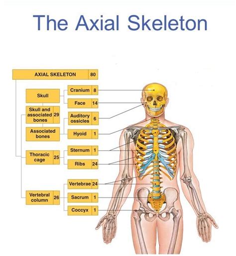
The axial skeleton forms the central axis of the body, acting as the core structural support. It's composed of 80 bones, meticulously arranged to protect vital organs and provide a foundation for the attachment of muscles involved in posture, breathing, and head and neck movement. Let's examine its key components:
1. The Skull: Protecting the Brain and Sensory Organs
The skull, arguably the most recognizable part of the axial skeleton, is a complex structure comprising 22 bones. It's divided into two main parts:
-
Cranium: This bony vault protects the brain, a vital organ responsible for virtually all bodily functions. The eight cranial bones – frontal, parietal (2), temporal (2), occipital, sphenoid, and ethmoid – are intricately fused together to create a strong, protective enclosure. Sutures, fibrous joints between these bones, allow for some flexibility during birth and growth but become largely immobile in adulthood.
-
Facial Bones: These fourteen bones form the framework of the face, providing support for the eyes, nose, and mouth. They also play crucial roles in chewing, speaking, and facial expression. Key facial bones include the maxillae (upper jaw), mandible (lower jaw – the only movable bone in the skull), zygomatic bones (cheekbones), and nasal bones.
2. The Vertebral Column: Providing Flexibility and Support
The vertebral column, or spine, is a flexible yet incredibly strong structure extending from the skull to the pelvis. It consists of 33 vertebrae, categorized into five regions:
-
Cervical Vertebrae (7): These are the neck vertebrae, providing support and flexibility for head movement. The first two, the atlas (C1) and axis (C2), have unique structures facilitating the nodding and rotation of the head.
-
Thoracic Vertebrae (12): These vertebrae articulate with the ribs, forming the rib cage. They are larger and more robust than cervical vertebrae, reflecting their role in supporting the weight of the upper body.
-
Lumbar Vertebrae (5): These are the largest and strongest vertebrae, supporting the weight of the upper body and facilitating bending and twisting movements. They are located in the lower back region.
-
Sacrum (1 fused bone): The sacrum is formed by the fusion of five sacral vertebrae. It's a wedge-shaped bone that connects the vertebral column to the pelvic girdle.
-
Coccyx (1 fused bone): The coccyx, or tailbone, is the most distal part of the vertebral column, formed by the fusion of three to five coccygeal vertebrae. It is a vestigial structure with limited functional importance in humans.
3. The Rib Cage: Protecting Vital Organs
The rib cage, also known as the thoracic cage, is a bony structure formed by 12 pairs of ribs, the sternum (breastbone), and costal cartilages. It protects vital organs within the thorax, including the heart and lungs. The ribs connect to the thoracic vertebrae posteriorly and either directly or indirectly to the sternum anteriorly.
-
True Ribs (1-7): These ribs connect directly to the sternum via their own costal cartilages.
-
False Ribs (8-10): These ribs connect indirectly to the sternum through the costal cartilage of the seventh rib.
-
Floating Ribs (11-12): These ribs have no anterior connection to the sternum.
The Appendicular Skeleton: Enabling Movement and Manipulation
The appendicular skeleton comprises the bones of the limbs and their supporting structures. It consists of approximately 126 bones and is responsible for facilitating movement, manipulation of objects, and locomotion. It's divided into the bones of the upper and lower limbs and the girdles connecting them to the axial skeleton.
1. The Pectoral (Shoulder) Girdle: Connecting the Upper Limbs
The pectoral girdle connects the upper limbs to the axial skeleton. It comprises two clavicles (collarbones) and two scapulae (shoulder blades). These bones provide a relatively flexible connection, allowing for a wide range of upper limb movement. The glenohumeral joint, where the humerus (upper arm bone) meets the scapula, is a ball-and-socket joint, contributing to the shoulder’s remarkable mobility.
2. The Upper Limbs: Fine Motor Skills and Strength
The upper limbs are designed for both fine motor skills and strength. Each upper limb consists of:
-
Humerus: The long bone of the upper arm.
-
Radius and Ulna: Two long bones in the forearm. The radius is the lateral bone (thumb side), and the ulna is the medial bone (pinky finger side). These bones rotate around each other, allowing for pronation (palm down) and supination (palm up).
-
Carpals: Eight small bones forming the wrist.
-
Metacarpals: Five long bones forming the palm.
-
Phalanges: Fourteen bones forming the fingers (three in each finger except the thumb, which has two).
3. The Pelvic (Hip) Girdle: Supporting the Lower Limbs
The pelvic girdle is a robust structure formed by two hip bones (coxal bones). Each coxal bone is formed by the fusion of three bones: the ilium, ischium, and pubis. The pelvic girdle provides strong support for the lower limbs and protects the pelvic organs. The sacroiliac joints connect the pelvic girdle to the sacrum, providing stability while still allowing for some movement.
4. The Lower Limbs: Locomotion and Weight Bearing
The lower limbs are adapted for weight-bearing and locomotion. Each lower limb consists of:
-
Femur: The longest and strongest bone in the body, located in the thigh.
-
Patella: The kneecap, a sesamoid bone embedded in the quadriceps tendon.
-
Tibia and Fibula: Two bones in the lower leg. The tibia (shinbone) is the weight-bearing bone, while the fibula provides lateral stability.
-
Tarsals: Seven bones forming the ankle.
-
Metatarsals: Five long bones forming the sole of the foot.
-
Phalanges: Fourteen bones forming the toes (three in each toe except the big toe, which has two).
Interconnections and Functional Significance
The axial and appendicular skeletons are not isolated entities; they are intricately interconnected, working together seamlessly to facilitate movement, maintain posture, and protect vital organs. The girdles – pectoral and pelvic – serve as crucial bridges, anchoring the appendicular skeleton to the axial skeleton. This structural integration allows for efficient transfer of forces during movement. For example, the force generated by leg muscles during walking is transmitted through the pelvic girdle, vertebral column, and ultimately to the ground.
The muscular system interacts extensively with the skeletal system. Skeletal muscles attach to bones via tendons, allowing for movement by pulling on the bones. The arrangement of bones and joints dictates the range and type of movement possible at each joint. For instance, the ball-and-socket joint of the shoulder allows for a greater range of motion than the hinge joint of the elbow.
The skeletal system also plays a critical role in hematopoiesis, the process of blood cell formation. Red bone marrow, found within certain bones, is the site of red blood cell, white blood cell, and platelet production. Furthermore, the skeletal system acts as a reservoir for essential minerals, primarily calcium and phosphorus. These minerals are constantly exchanged between the bones and the bloodstream, maintaining blood mineral homeostasis.
Conclusion
The axial and appendicular skeletons, although distinct divisions, form a unified and remarkably efficient system. Their interconnected structure and intricate interactions with the muscular and circulatory systems highlight the sophistication and importance of the human skeletal system. Understanding the specific roles of each bone and the overall functional integration of these two divisions is essential for appreciating the complex mechanics of human movement, posture, and overall bodily function. Further exploration into the microscopic structure of bone tissue, the specifics of joint articulation, and the intricate interplay between the skeletal system and other organ systems would enrich this understanding further.
Latest Posts
Latest Posts
-
Which Elements Have The Highest Ionization Energy
Apr 06, 2025
-
Calculate Total Magnification Of A Microscope
Apr 06, 2025
-
What Are The Resources Of A Business
Apr 06, 2025
-
Label The Bones Of The Appendicular Skeleton
Apr 06, 2025
-
The Carbohydrates Glucose Galactose And Fructose
Apr 06, 2025
Related Post
Thank you for visiting our website which covers about What Are The Two Major Divisions Of The Skeletal System . We hope the information provided has been useful to you. Feel free to contact us if you have any questions or need further assistance. See you next time and don't miss to bookmark.
