What Are Two Divisions Of The Skeleton
Muz Play
Mar 31, 2025 · 6 min read
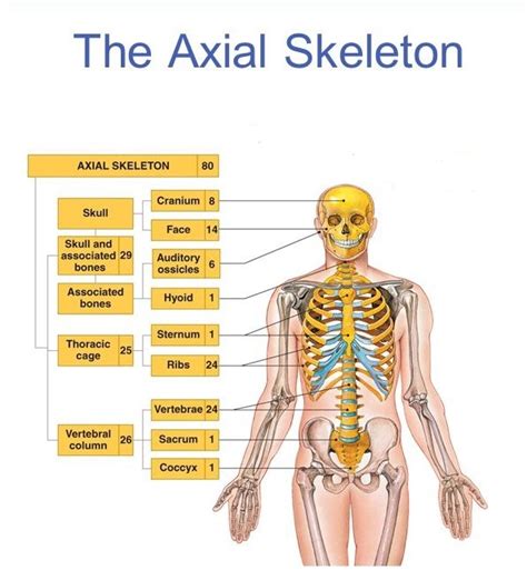
Table of Contents
What Are the Two Divisions of the Skeleton? A Deep Dive into Axial and Appendicular Anatomy
The human skeleton, a marvel of biological engineering, provides structural support, protects vital organs, and facilitates movement. Understanding its structure is crucial for anyone interested in anatomy, physiology, or related fields. This comprehensive guide delves into the two main divisions of the skeleton: the axial skeleton and the appendicular skeleton. We'll explore their components, functions, and interconnectedness, providing a detailed overview for both beginners and those seeking a refresher on this fundamental aspect of human biology.
The Axial Skeleton: The Body's Central Support Structure
The axial skeleton forms the central axis of the body, providing the fundamental framework for the head, neck, and trunk. Think of it as the core, the foundational structure upon which the rest of the skeleton is built. It comprises approximately 80 bones, and its primary functions include:
- Protecting vital organs: The skull encases the brain, the vertebrae safeguard the spinal cord, and the rib cage shields the heart and lungs. This protective role is paramount for survival.
- Supporting the body's weight: The vertebral column, a critical part of the axial skeleton, bears the weight of the upper body and transmits it to the lower limbs.
- Providing attachment points for muscles: Many muscles responsible for movement and posture attach to the bones of the axial skeleton, enabling various actions.
Components of the Axial Skeleton: A Detailed Look
Let's break down the axial skeleton into its key components:
-
The Skull (Cranium and Facial Bones): The skull is arguably the most recognizable part of the axial skeleton. The cranium protects the brain, while the facial bones contribute to facial structure and sensory functions (sight, smell, taste). Within the skull are numerous bones, including the frontal bone, parietal bones, temporal bones, occipital bone, sphenoid bone, ethmoid bone, and various facial bones like the nasal bones, zygomatic bones, maxilla, and mandible (jawbone). The intricate structure of the skull allows for protection while providing openings for sensory organs and passageways for blood vessels and nerves.
-
The Vertebral Column (Spine): This crucial structure consists of 33 vertebrae, grouped into five regions:
- Cervical Vertebrae (C1-C7): The seven cervical vertebrae in the neck are characterized by their small size and unique features, like the atlas (C1) and axis (C2) which allow for head rotation and flexion.
- Thoracic Vertebrae (T1-T12): Twelve thoracic vertebrae articulate with the ribs, forming the posterior part of the rib cage.
- Lumbar Vertebrae (L1-L5): These five lumbar vertebrae in the lower back are the largest and strongest, supporting the majority of the upper body's weight.
- Sacrum: This triangular bone is formed from the fusion of five sacral vertebrae and articulates with the hip bones.
- Coccyx (Tailbone): This small, vestigial structure is formed from the fusion of three to five coccygeal vertebrae.
-
The Rib Cage (Thoracic Cage): This bony cage protects the heart, lungs, and other vital organs in the chest. It consists of 12 pairs of ribs, each articulating with a thoracic vertebra posteriorly. The first seven pairs are true ribs, attaching directly to the sternum (breastbone). Ribs 8-10 are false ribs, indirectly attached to the sternum via cartilage. Ribs 11 and 12 are floating ribs, lacking any sternal attachment. The sternum itself is a flat bone consisting of three parts: the manubrium, body, and xiphoid process.
The Appendicular Skeleton: The Skeleton of Movement and Manipulation
The appendicular skeleton comprises the bones of the limbs (arms and legs) and their supporting structures (girdles). It's the part of the skeleton responsible for locomotion and manipulation of objects. This division contains approximately 126 bones and is vital for mobility and interaction with the environment.
Components of the Appendicular Skeleton: A Closer Examination
-
The Pectoral Girdle (Shoulder Girdle): This girdle connects the upper limbs to the axial skeleton. It consists of two clavicles (collarbones) and two scapulae (shoulder blades). The relatively loose attachment of the pectoral girdle allows for a wide range of arm movements.
-
The Upper Limbs: Each upper limb has 30 bones, including:
- Humerus: The long bone of the upper arm.
- Radius and Ulna: The two bones of the forearm, allowing for pronation and supination (rotation of the hand).
- Carpals: Eight small bones forming the wrist.
- Metacarpals: Five bones forming the palm.
- Phalanges: Fourteen bones forming the fingers (three in each finger except the thumb, which has two).
-
The Pelvic Girdle (Hip Girdle): This robust girdle connects the lower limbs to the axial skeleton. It's formed by two hip bones (ossa coxae), each composed of three fused bones: the ilium, ischium, and pubis. The pelvic girdle provides strong support for the trunk and protects the pelvic organs. The structure and size of the pelvic girdle differ significantly between males and females.
-
The Lower Limbs: Each lower limb also contains 30 bones:
- Femur: The longest and strongest bone in the body, located in the thigh.
- Patella (Kneecap): A sesamoid bone embedded in the quadriceps tendon, protecting the knee joint.
- Tibia and Fibula: The two bones of the lower leg; the tibia is the weight-bearing bone.
- Tarsals: Seven bones forming the ankle.
- Metatarsals: Five bones forming the sole of the foot.
- Phalanges: Fourteen bones forming the toes (three in each toe except the big toe, which has two).
The Interplay Between Axial and Appendicular Skeletons
The axial and appendicular skeletons are not isolated entities; they function together as a unified system. The appendicular skeleton relies on the axial skeleton for support and stability. The girdles – pectoral and pelvic – act as crucial bridges, connecting the limbs to the axial skeleton. This integration allows for coordinated movement, maintaining balance and posture. Muscle attachments spanning both skeletal divisions further emphasize their interconnectedness.
Clinical Significance: Disorders Affecting the Skeleton
Numerous disorders can affect both the axial and appendicular skeletons, highlighting the importance of skeletal health. Some examples include:
- Scoliosis: A lateral curvature of the spine, primarily affecting the axial skeleton.
- Osteoporosis: A condition characterized by decreased bone density, weakening bones throughout the entire skeleton.
- Fractures: Bones can fracture due to trauma or underlying conditions like osteoporosis, affecting both divisions.
- Osteoarthritis: Degenerative joint disease that commonly affects weight-bearing joints in the appendicular skeleton, such as the knees and hips.
- Spina Bifida: A birth defect affecting the development of the spinal cord and vertebrae, impacting the axial skeleton.
Conclusion: A Comprehensive Understanding of Skeletal Structure
This detailed exploration of the axial and appendicular skeletons underscores their critical roles in supporting the body, protecting vital organs, and enabling movement. Understanding the specific bones, their arrangement, and their interrelationships provides a strong foundation for appreciating the complexity and beauty of the human body. Furthermore, recognizing the potential disorders impacting the skeleton emphasizes the importance of maintaining skeletal health through proper nutrition, exercise, and preventative care. From protecting vital organs to facilitating graceful movement, the skeleton's two main divisions work in perfect harmony, showcasing the remarkable design of the human form. Further research into specific bones and their functions can provide an even deeper appreciation for this intricate and essential part of human anatomy.
Latest Posts
Latest Posts
-
What Is The Monomer Of A Dna Molecule
Apr 01, 2025
-
When Does The Segregation Of Alleles Occur
Apr 01, 2025
-
Cardiac Muscles Differ From Skeletal Muscles In That They
Apr 01, 2025
-
Closing Entries Are Journalized And Posted
Apr 01, 2025
-
How To Calculate Parts Per Thousand
Apr 01, 2025
Related Post
Thank you for visiting our website which covers about What Are Two Divisions Of The Skeleton . We hope the information provided has been useful to you. Feel free to contact us if you have any questions or need further assistance. See you next time and don't miss to bookmark.
