What Color Is An Animal Cell Membrane
Muz Play
Apr 05, 2025 · 6 min read

Table of Contents
What Color Is an Animal Cell Membrane? The Surprising Answer and the Science Behind It
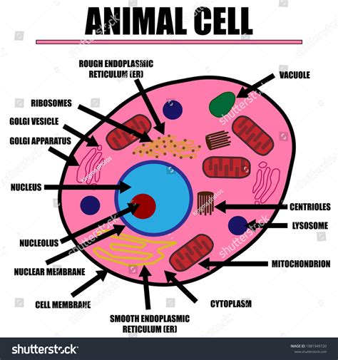
The question, "What color is an animal cell membrane?" might seem simple at first glance. However, the answer is far more nuanced than a simple color designation. Understanding the true nature of the cell membrane requires delving into its intricate structure and function, revealing a fascinating story of molecular organization and dynamic interactions. This article will explore the reasons why assigning a specific color to the cell membrane is misleading and will discuss the fascinating science behind this fundamental component of life.
The Illusion of Color: Why You Won't See a Colored Membrane
The short answer is: animal cell membranes are colorless. Or, more accurately, they are transparent or translucent. This lack of color is directly related to their structure and composition. Unlike pigments that absorb specific wavelengths of light, creating the colors we see, the cell membrane's components primarily scatter and transmit light.
We're used to associating color with macroscopic objects. However, at the microscopic level, things are different. The cell membrane is incredibly thin – approximately 7-10 nanometers. This is far thinner than the wavelength of visible light, making it effectively invisible to the naked eye. Even under a standard light microscope, the membrane itself is often indistinguishable from the surrounding cytoplasm.
The Structure of the Animal Cell Membrane: A Closer Look
To understand why the cell membrane lacks a discernible color, we need to examine its structure. The fluid mosaic model is the currently accepted model describing its architecture. This model highlights the key components:
-
Phospholipid Bilayer: This forms the basic framework of the membrane. Phospholipids are amphipathic molecules, meaning they have both hydrophilic (water-loving) and hydrophobic (water-fearing) regions. The hydrophilic heads face outward, interacting with the aqueous environments inside and outside the cell, while the hydrophobic tails cluster together in the interior of the membrane. This bilayer structure is crucial for maintaining the cell's integrity and selectively controlling the passage of substances. It's this colorless phospholipid bilayer that forms the structural basis, creating transparency rather than color.
-
Proteins: Embedded within the phospholipid bilayer are a variety of proteins. These proteins serve numerous functions, including:
- Transport Proteins: Facilitate the movement of molecules across the membrane.
- Receptor Proteins: Bind to signaling molecules, triggering cellular responses.
- Enzymes: Catalyze biochemical reactions within the membrane.
- Structural Proteins: Maintain the integrity and shape of the membrane.
These proteins, while vital, are also generally colorless or transparent at the individual level and do not contribute to a discernible color when combined within the membrane.
-
Carbohydrates: These are attached to both lipids (glycolipids) and proteins (glycoproteins) on the outer surface of the membrane. They play roles in cell recognition, adhesion, and communication. Again, these carbohydrate components do not impart any significant color to the membrane.
-
Cholesterol: This lipid molecule is interspersed among the phospholipids, influencing membrane fluidity and stability. Like the other components, cholesterol does not contribute to any noticeable coloration.
Advanced Microscopy Techniques: Visualizing the Membrane
While the cell membrane itself isn't inherently colored, advanced microscopy techniques can allow us to visualize its structure and components.
-
Electron Microscopy: This powerful technique provides high-resolution images of the cell membrane, revealing its layered structure and the distribution of proteins. However, electron microscopy uses electron beams, not visible light, so the images are black and white, not representative of actual color. Color is often added artificially to highlight specific structures in the processed images.
-
Fluorescence Microscopy: This technique uses fluorescent dyes to label specific components of the cell, allowing researchers to visualize their localization and dynamics. For example, fluorescent antibodies can be used to label specific membrane proteins. While this provides stunning visual representations, the color observed is due to the fluorescent dye, not the inherent color of the membrane itself. Different dyes are selected to label specific target molecules and therefore different colors are observed depending on the target molecule.
-
Confocal Microscopy: This advanced technique improves the resolution and clarity of fluorescence microscopy, further enhancing our ability to study membrane components. Again, the color observed is from the fluorescent label and not the membrane itself.
Why the Color Question Matters: Understanding Cellular Processes
While the lack of color in the cell membrane might seem like a trivial detail, understanding its composition and structure is crucial to comprehend essential cellular processes:
-
Selective Permeability: The membrane's structure is paramount in controlling the movement of substances into and out of the cell. This selective permeability maintains a stable internal environment crucial for life. Understanding the interactions of molecules with the membrane's phospholipid bilayer and embedded proteins is crucial for understanding transport mechanisms such as active and passive transport, osmosis, and diffusion.
-
Cell Signaling: The membrane plays a vital role in cell signaling, the process of communication between cells. Receptor proteins on the membrane bind to signaling molecules, triggering intracellular cascades that regulate various cellular processes like growth, differentiation, and apoptosis. Understanding the spatial arrangement of these proteins and their interactions with signaling molecules are key to understanding how cells communicate with their environment.
-
Cell Adhesion: The interactions between cell surface molecules (glycoproteins and glycolipids) mediate cell adhesion, allowing cells to interact with each other and the extracellular matrix. This process is fundamental in tissue formation, development, and immune responses.
-
Membrane Dynamics: The cell membrane is a highly dynamic structure, constantly undergoing remodeling and reorganization. Understanding the fluidity of the membrane and the movement of its components is essential for understanding processes like endocytosis (cell uptake of substances) and exocytosis (cell secretion).
Beyond Animal Cells: Variations in Membrane Composition
It's important to note that the composition and structure of cell membranes can vary slightly across different cell types and even across different regions of a single cell. However, the fundamental principle remains the same: the membrane's core structure is a colorless phospholipid bilayer, and the various embedded proteins and carbohydrates do not impart any significant color. While some variations exist, the overarching transparency remains a consistent feature.
Conclusion: Transparency, Not Color, Defines the Cell Membrane
The question "What color is an animal cell membrane?" highlights a crucial point: the limitations of our visual perception at the microscopic level. While the absence of inherent color might seem unremarkable, it reflects the fundamental nature of the cell membrane – a transparent, dynamic structure essential for life. A deeper understanding of the membrane's colorless, intricate structure is vital for grasping its multifaceted role in cellular processes and overall biological function. The colorless nature of the cell membrane is not a lack of significance but a reflection of its elegant and efficient design, optimized for the crucial tasks of selective permeability, cell signaling, and maintaining cellular integrity. Further research into the intricate details of membrane structure and function will undoubtedly continue to reveal more fascinating aspects of this essential component of life.
Latest Posts
Latest Posts
-
Formula For Kinetic Energy Of A Spring
Apr 06, 2025
-
What Are The Three Types Of Asexual Reproduction
Apr 06, 2025
-
A Measure Of Quantity Of Matter Is
Apr 06, 2025
-
Electric Field Lines Between Two Positive Charges
Apr 06, 2025
-
Which Compounds Will Dissolve In Water
Apr 06, 2025
Related Post
Thank you for visiting our website which covers about What Color Is An Animal Cell Membrane . We hope the information provided has been useful to you. Feel free to contact us if you have any questions or need further assistance. See you next time and don't miss to bookmark.
