What Is A Compound Light Microscope
Muz Play
Mar 21, 2025 · 6 min read
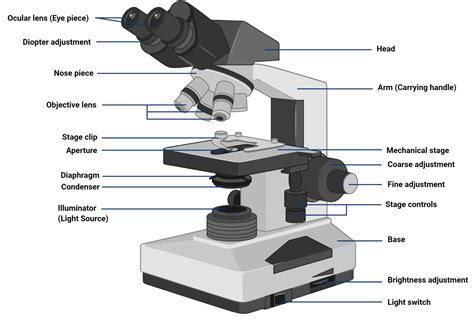
Table of Contents
What is a Compound Light Microscope? A Comprehensive Guide
The compound light microscope, a cornerstone of biological and medical research, allows us to visualize the intricate details of the microscopic world invisible to the naked eye. Understanding its components, functionalities, and applications is crucial for anyone working in fields requiring microscopic analysis. This comprehensive guide will delve into every aspect of the compound light microscope, from its basic principles to advanced techniques.
Understanding the Fundamentals: How it Works
The compound light microscope's name itself hints at its key feature: it uses a compound system of lenses to magnify the image of a specimen. Unlike a simple microscope with a single lens, the compound microscope employs two lens systems: the objective lens and the eyepiece lens (ocular lens). Each lens magnifies the image, resulting in a significantly higher total magnification than a simple microscope could achieve.
The process begins with the light source, typically located at the base of the microscope. This light passes upwards through a condenser lens, which focuses the light onto the specimen on the stage. The specimen, usually mounted on a glass slide, is illuminated. The light then passes through the specimen and into the objective lens, which produces a magnified real image. This real image is then further magnified by the eyepiece lens, producing a virtual image that the observer sees through the eyepiece.
Key Components of a Compound Light Microscope: A Detailed Breakdown
Let's explore the essential components in greater detail:
-
Eyepiece Lens (Ocular Lens): This is the lens you look through. It usually provides a magnification of 10x. Some microscopes have binocular eyepieces, allowing for comfortable viewing with both eyes.
-
Objective Lenses: These are the most crucial lenses, responsible for the initial magnification of the specimen. A typical compound microscope has multiple objective lenses with different magnification powers, commonly 4x, 10x, 40x, and 100x (oil immersion). The magnification of the objective lens is engraved on its barrel.
-
Revolving Nosepiece (Turret): This rotating mechanism holds the objective lenses, allowing for easy switching between different magnifications.
-
Stage: This is the flat platform where the microscope slide is placed. Many microscopes have stage clips to hold the slide securely in place. Advanced models include mechanical stage controls for precise movement of the slide.
-
Condenser Lens: Located below the stage, this lens focuses the light from the light source onto the specimen. Adjusting the condenser's height and diaphragm controls the intensity and evenness of illumination, crucial for optimal image quality.
-
Diaphragm: Part of the condenser, this adjusts the amount of light passing through the condenser, influencing contrast and brightness. A closed diaphragm increases contrast, while an open diaphragm increases brightness.
-
Light Source: This provides the illumination for the specimen. Modern microscopes often use LED light sources, offering bright, energy-efficient illumination.
-
Coarse Adjustment Knob: This large knob moves the stage up and down in large increments, allowing for quick focusing, particularly useful at lower magnifications.
-
Fine Adjustment Knob: This smaller knob makes precise adjustments to the focus, crucial for achieving sharp images at higher magnifications.
-
Arm: This supports the body tube and connects it to the base.
-
Base: The bottom of the microscope providing stability and support.
Calculating Total Magnification
The total magnification of the compound microscope is calculated by multiplying the magnification of the objective lens by the magnification of the eyepiece lens. For example, with a 10x eyepiece and a 40x objective lens, the total magnification is 400x (10 x 40 = 400).
Types of Compound Light Microscopes
While the basic principles remain consistent, variations exist in the design and capabilities of compound light microscopes. These include:
-
Monocular Microscopes: These have a single eyepiece.
-
Binocular Microscopes: These have two eyepieces, offering a more comfortable and less eye-straining viewing experience.
-
Trinocular Microscopes: These have two eyepieces and an additional port for attaching a camera, allowing for image capture and documentation.
-
Inverted Microscopes: In these microscopes, the light source is above the stage, and the objective lenses are below. This design is particularly useful for observing living cells in culture dishes.
-
Phase Contrast Microscopes: These microscopes enhance the contrast of transparent specimens, allowing for better visualization of cellular structures without the need for staining.
-
Darkfield Microscopes: These microscopes only illuminate light scattered by the specimen, creating a bright specimen against a dark background. This is useful for observing unstained, transparent specimens.
-
Fluorescence Microscopes: These microscopes use fluorescent dyes or proteins to label specific structures within the specimen, allowing for highly specific visualization. They are widely used in various biological and medical applications.
Preparing Specimens for Microscopy: A Crucial Step
Proper specimen preparation is critical for obtaining high-quality images. This involves several steps, including:
-
Sample Collection: Obtaining a representative sample is the first step.
-
Sample Preparation: This depends on the nature of the specimen and the type of microscopy being used. Techniques include staining, fixation, and sectioning.
-
Mounting: The prepared specimen is then mounted onto a glass microscope slide, often using a mounting medium to prevent drying and preserve the specimen.
-
Coverslip Application: A coverslip is carefully placed over the specimen to protect it and ensure even illumination.
Applications of Compound Light Microscopes: A Wide Range of Uses
The compound light microscope's versatility makes it an indispensable tool across numerous disciplines:
-
Biology: Studying cells, tissues, microorganisms, and other biological specimens.
-
Medicine: Diagnosing diseases, analyzing blood samples, examining tissue biopsies.
-
Pathology: Identifying pathogens and cancerous cells.
-
Histology: Examining the microscopic structure of tissues.
-
Cytology: Studying individual cells.
-
Environmental Science: Analyzing water samples, identifying pollutants, observing microorganisms.
-
Education: Teaching basic biological principles and microscopy techniques.
Advanced Techniques and Considerations
Beyond the basic usage, several advanced techniques further enhance the capabilities of the compound light microscope:
-
Oil Immersion Microscopy: Using immersion oil with the 100x objective lens improves resolution by reducing light refraction.
-
Microphotography: Capturing images of microscopic specimens using a camera attached to the microscope.
-
Digital Microscopy: Utilizing digital cameras and software for image capture, analysis, and storage.
-
Fluorescence Microscopy: Observing fluorescently labeled specimens.
-
Confocal Microscopy: A more advanced technique that uses lasers to create high-resolution images of thick specimens.
Maintenance and Care of Your Compound Light Microscope: Ensuring Longevity
Proper maintenance ensures the longevity and accuracy of your compound light microscope:
-
Cleanliness: Regularly clean the lenses with lens paper and lens cleaning solution. Avoid touching the lenses with your fingers.
-
Storage: Store the microscope in a clean, dry, and dust-free environment.
-
Handling: Handle the microscope with care, avoiding sudden movements or impacts.
-
Calibration: Periodically check the calibration of the microscope to ensure accurate measurements.
Conclusion: An Indispensable Tool for Exploration
The compound light microscope remains a fundamental instrument in numerous scientific disciplines. Its ability to reveal the intricate details of the microscopic world has revolutionized our understanding of biology, medicine, and many other fields. By understanding its principles, components, and applications, we can effectively harness its power for exploration and discovery. This guide provides a solid foundation for anyone seeking to understand and utilize this powerful tool. Further exploration into specific applications and advanced techniques will undoubtedly enhance your understanding and proficiency in microscopy.
Latest Posts
Latest Posts
-
Song Lyrics Ode To Billy Joe
Mar 27, 2025
-
What Happens During The Reduction Stage Of The Calvin Cycle
Mar 27, 2025
-
Is Solid To Liquid Endothermic Or Exothermic
Mar 27, 2025
-
What Does A Negative Enthalpy Mean
Mar 27, 2025
-
Divides The Body Into Anterior And Posterior Portions
Mar 27, 2025
Related Post
Thank you for visiting our website which covers about What Is A Compound Light Microscope . We hope the information provided has been useful to you. Feel free to contact us if you have any questions or need further assistance. See you next time and don't miss to bookmark.
