What Is In The Cranial Cavity
Muz Play
Apr 01, 2025 · 6 min read
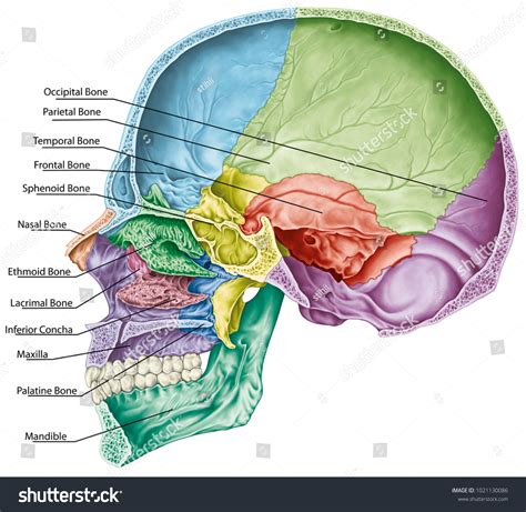
Table of Contents
What's Inside the Cranial Cavity? A Comprehensive Guide
The human skull, a marvel of biological engineering, protects the delicate and vital contents of the cranial cavity. Understanding the structures within this bony fortress is crucial for comprehending neurological function, diagnosing diseases, and appreciating the complexity of the human brain. This comprehensive guide delves into the intricate anatomy of the cranial cavity, exploring its major components and their interrelationships.
The Bony Structure: Protecting the Brain
Before examining the contents, let's consider the protective shell itself. The cranial cavity is formed by the cranial bones: the frontal bone, two parietal bones, two temporal bones, the occipital bone, the sphenoid bone, and the ethmoid bone. These bones are intricately joined by sutures, strong fibrous joints that provide stability while allowing for some flexibility during birth and growth. The shape of the cranial cavity is not uniform; it presents various fossae (depressions) and foramina (openings) designed to accommodate the brain and its associated structures.
Key Features of the Cranial Bones:
- Frontal Bone: Forms the forehead and the anterior portion of the cranial base. Houses the frontal sinuses.
- Parietal Bones: Form the superior and lateral aspects of the skull.
- Temporal Bones: Located on either side of the skull, these bones house the organs of hearing and balance. They contain the external auditory meatus, mastoid process, and the mandibular fossa (where the lower jaw articulates).
- Occipital Bone: Forms the posterior portion of the skull and the base of the skull. Contains the foramen magnum, a large opening through which the spinal cord passes.
- Sphenoid Bone: A complex, bat-shaped bone located in the middle of the cranial base. Contains the sella turcica, which houses the pituitary gland.
- Ethmoid Bone: Located anterior to the sphenoid bone, it forms part of the nasal cavity and contributes to the orbits (eye sockets). It contains the cribriform plate, through which olfactory nerves pass.
These bones work in concert to create a robust, yet lightweight, protective structure for the brain and its associated structures. The intricate arrangement of sutures ensures both strength and flexibility.
The Brain: The Master Control Center
The most significant occupant of the cranial cavity is undoubtedly the brain. This complex organ is responsible for virtually all aspects of our conscious and unconscious functions, from higher-level thought processes to basic life-sustaining activities. The brain is broadly divided into three major parts: the cerebrum, cerebellum, and brainstem.
The Cerebrum: The Seat of Higher Cognition
The cerebrum is the largest part of the brain, occupying most of the cranial cavity. It's divided into two hemispheres (right and left), each further subdivided into four lobes: frontal, parietal, temporal, and occipital.
- Frontal Lobe: Responsible for higher-order cognitive functions, including planning, decision-making, voluntary movement, and speech production.
- Parietal Lobe: Processes sensory information from the body, including touch, temperature, pain, and spatial awareness.
- Temporal Lobe: Involved in auditory processing, memory formation, and language comprehension.
- Occipital Lobe: Primarily responsible for visual processing.
The cerebrum's surface is highly convoluted, characterized by gyri (ridges) and sulci (grooves). This intricate folding maximizes the surface area available for neuronal connections, enhancing cognitive capabilities. Deep within the cerebrum lie the basal ganglia, crucial for motor control and coordination. The corpus callosum, a thick band of nerve fibers, connects the two cerebral hemispheres, facilitating communication between them.
The Cerebellum: The Master of Motor Control
Located beneath the cerebrum at the back of the skull, the cerebellum is crucial for coordinating movement, balance, and posture. It receives input from various parts of the brain and body and fine-tunes motor commands to ensure smooth, coordinated movements. Damage to the cerebellum can result in ataxia (loss of coordination) and tremors.
The Brainstem: Connecting the Brain and Spinal Cord
The brainstem connects the cerebrum and cerebellum to the spinal cord. It consists of the midbrain, pons, and medulla oblongata. These structures control vital life functions such as breathing, heart rate, and blood pressure. The brainstem also plays a crucial role in regulating sleep-wake cycles and consciousness. Cranial nerves III-XII originate from the brainstem.
Meninges: Protective Layers Surrounding the Brain
The brain is not directly in contact with the bony skull. Three layers of protective membranes, called meninges, separate the brain from the skull:
- Dura Mater: The outermost and toughest layer, it is a thick, fibrous membrane that adheres to the inner surface of the skull. It has two layers – the periosteal and meningeal layers. Between these layers are the dural venous sinuses, which collect venous blood from the brain. The dura mater also forms folds that separate different parts of the brain.
- Arachnoid Mater: The middle layer, it is a delicate, web-like membrane that lies beneath the dura mater. The subarachnoid space, located between the arachnoid and pia mater, contains cerebrospinal fluid (CSF).
- Pia Mater: The innermost layer, it is a thin, transparent membrane that closely adheres to the surface of the brain. It follows the contours of the brain's gyri and sulci.
Cerebrospinal Fluid (CSF): A Protective Fluid
CSF is a clear, colorless fluid that circulates within the subarachnoid space, ventricles (cavities within the brain), and spinal canal. It provides buoyancy, cushioning the brain against impact and reducing pressure on the brain tissue. CSF also plays a role in removing metabolic waste products from the brain and maintaining a stable chemical environment.
Blood Vessels: Nourishing the Brain
The brain's high metabolic demands require a constant supply of oxygen and nutrients. A vast network of blood vessels, including arteries and veins, supplies the brain with blood. The major arteries supplying the brain include the internal carotid arteries and the vertebral arteries. Venous drainage is accomplished by a system of dural venous sinuses and veins that ultimately drain into the jugular veins. The circle of Willis, a critical anastomosis of arteries at the base of the brain, provides redundancy in blood supply, minimizing the effects of vessel occlusion.
Cranial Nerves: Connecting the Brain to the Peripheral Nervous System
Twelve pairs of cranial nerves emerge from the brainstem and pass through foramina in the skull to innervate various parts of the head, neck, and torso. These nerves control functions such as vision, hearing, taste, smell, facial expression, swallowing, and head and shoulder movement.
Other Structures Within the Cranial Cavity:
While the brain is the dominant structure, other important components reside within the cranial cavity:
- Pituitary Gland: Located in the sella turcica of the sphenoid bone, this small endocrine gland produces numerous hormones that regulate various bodily functions.
- Hypothalamus: A small region of the brain located below the thalamus, it plays a crucial role in regulating homeostasis, including body temperature, hunger, thirst, and sleep cycles.
- Pineal Gland: Located near the center of the brain, it produces melatonin, a hormone that regulates sleep-wake cycles.
- Blood Vessels and Nerves: A complex network of arteries, veins, and nerves provide blood supply and innervation to the brain and other structures within the cranial cavity.
Conclusion: A Complex and Vital Space
The cranial cavity is a remarkably complex space containing a multitude of vital structures. The intricate interplay between the bony skull, meninges, cerebrospinal fluid, blood vessels, and neural tissues ensures the proper functioning of the brain, the master control center of the human body. A thorough understanding of the cranial cavity's anatomy is fundamental to various medical fields, enabling accurate diagnosis and treatment of neurological disorders and injuries. Further exploration into the specific functions of each structure within the cranial cavity will undoubtedly deepen your appreciation for the wonders of human biology.
Latest Posts
Latest Posts
-
Thesis Statement For Narrative Essay Example
Apr 02, 2025
-
Inborn Nonspecific Defenses Include And Barriers
Apr 02, 2025
-
Jewish Murals From The First Century Ce Depict
Apr 02, 2025
-
Which Quadratic Function Is Represented By The Graph
Apr 02, 2025
-
An Organized Arrangement Of Elements According To Their Atomic Number
Apr 02, 2025
Related Post
Thank you for visiting our website which covers about What Is In The Cranial Cavity . We hope the information provided has been useful to you. Feel free to contact us if you have any questions or need further assistance. See you next time and don't miss to bookmark.
