What Is The Color Of A Nucleus
Muz Play
Mar 23, 2025 · 5 min read
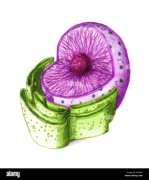
Table of Contents
What is the Color of a Nucleus? A Deep Dive into Cellular Structures and Microscopy
The question, "What is the color of a nucleus?" might seem deceptively simple. However, the answer is far more nuanced than a single color. The apparent color of a nucleus depends heavily on several factors, including the type of cell, the staining techniques used, and the method of observation (light microscopy versus electron microscopy). This exploration will delve into the intricacies of nuclear structure, microscopy techniques, and the reasons behind the varied appearances of the nucleus under different conditions.
The Nucleus: The Control Center of the Cell
Before discussing color, it's crucial to understand the nucleus itself. This organelle, a defining feature of eukaryotic cells (plants, animals, fungi, and protists), houses the cell's genetic material—DNA—organized into chromosomes. The nucleus isn't a uniformly colored blob; rather, it's a complex structure with several components:
Nuclear Envelope:
The nucleus is bounded by a double membrane known as the nuclear envelope. This envelope regulates the transport of molecules in and out of the nucleus, maintaining a distinct internal environment. This membrane itself is essentially colorless, though it's studded with nuclear pores.
Chromatin:
Within the nucleus resides chromatin, a complex of DNA and proteins. Chromatin is not inherently colored; its appearance is heavily influenced by staining techniques used in microscopy. During cell division, chromatin condenses into distinct chromosomes, structures readily visible even without intense staining.
Nucleolus:
The nucleolus, a prominent, dense structure within the nucleus, is the site of ribosome biogenesis. Like chromatin, its color is not inherent but depends on staining and microscopy.
Microscopy and Staining: Unveiling the Nucleus's Apparent Color
The perceived color of the nucleus is almost entirely a product of microscopy and staining techniques. The naked eye cannot see the nucleus. Microscopy allows us to visualize this crucial organelle, but even then, its color is not intrinsic.
Light Microscopy:
Light microscopy, the most common technique for visualizing cells, uses visible light to illuminate the sample. Without staining, the nucleus may appear slightly translucent or grayish, barely distinguishable from the surrounding cytoplasm. The use of stains, however, dramatically alters its appearance:
-
Hematoxylin and Eosin (H&E) Staining: This widely used staining technique in histology, employs hematoxylin, a basic dye that stains acidic structures such as DNA a deep purple or blue. Eosin, an acidic dye, stains other cellular components like cytoplasm pink or red. Thus, in H&E stained preparations, the nucleus typically appears deep purple or blue.
-
Other Stains: Various other stains target specific components of the nucleus. For example, some fluorescent dyes can be used to specifically label DNA, leading to a brightly fluorescent nucleus, depending on the dye used, ranging from green to red to blue. The choice of stain dictates the perceived color.
Electron Microscopy:
Electron microscopy offers far higher resolution than light microscopy, allowing visualization of cellular structures at the nanometer scale. Electron microscopy doesn't use stains in the same way as light microscopy; it relies on electron scattering to create images. Consequently, the nucleus isn't colored in the traditional sense but appears as variations in grayscale, depending on the density of different nuclear components. Dense regions like the nucleolus appear darker, while less dense areas appear lighter.
Factors Influencing Nuclear Appearance: Beyond Staining
The apparent color of a nucleus, even with staining, isn't always uniform. Several factors contribute to variations in appearance:
Cell Type:
The size and structure of the nucleus vary between different cell types. Neurons, for example, often have large, irregularly shaped nuclei, while red blood cells lack a nucleus altogether. These differences can subtly influence the perceived color and intensity of staining.
Cell Cycle Stage:
The nucleus's appearance changes dramatically throughout the cell cycle. During interphase (the period between cell divisions), chromatin is relatively diffuse, and the nucleus appears relatively uniform. During mitosis (cell division), chromatin condenses into highly compact chromosomes, making the nucleus appear more structured and potentially altering the staining intensity.
Cellular Health:
The health of a cell can affect the nucleus's appearance. Damaged or stressed cells may exhibit changes in nuclear morphology (shape and structure), including changes in chromatin organization and condensation, which might alter the way the nucleus takes up stain.
Specimen Preparation:
The process of preparing a sample for microscopy can also influence the appearance of the nucleus. Techniques like fixation and embedding can introduce artifacts that might affect staining and the overall image.
The Importance of Understanding Nuclear Color in Research
Understanding the color of a nucleus and the factors that influence it is critical in various research areas:
-
Histology and Pathology: The appearance of the nucleus in stained tissue sections is fundamental to diagnosing diseases. Abnormal nuclear morphology, such as changes in size, shape, and staining intensity, is a key indicator of various cancers and other pathologies.
-
Cell Biology: Studying the nucleus's structure and function requires precise visualization techniques. The choice of stains and microscopy methods are crucial for obtaining meaningful data on nuclear processes like gene expression and DNA replication.
-
Genetic Engineering and Genomics: Observing and manipulating the nucleus is essential in genetic engineering and genomics research. Understanding how different dyes interact with the nucleus helps researchers track gene expression, identify mutations, and conduct other crucial experiments.
Conclusion: No Single Answer
There's no single definitive answer to the question, "What is the color of a nucleus?" The answer is contingent on a number of factors, primarily the staining technique used and the type of microscopy employed. In H&E stained preparations viewed under light microscopy, the nucleus typically appears deep purple or blue. However, using different dyes or electron microscopy will yield different appearances. The complexity and variability of the nucleus’s appearance highlight the sophistication of cellular structures and the importance of choosing the appropriate techniques for effective visualization and analysis. Understanding these complexities is crucial for researchers in various biological fields, from clinical diagnostics to fundamental cell biology. The next time you see a diagram of a cell, remember the fascinating story behind the seemingly simple color of its nucleus.
Latest Posts
Latest Posts
-
Separation Of Variables Partial Differential Equations
Mar 24, 2025
-
How To Calculate Bond Dissociation Energy
Mar 24, 2025
-
Which Of The Following Is A Chemical Property Of Matter
Mar 24, 2025
-
Formula For Motion With Constant Acceleration
Mar 24, 2025
-
How Do You Calculate Rate Of Diffusion
Mar 24, 2025
Related Post
Thank you for visiting our website which covers about What Is The Color Of A Nucleus . We hope the information provided has been useful to you. Feel free to contact us if you have any questions or need further assistance. See you next time and don't miss to bookmark.
