What Is The Structural And Functional Unit Of The Kidney
Muz Play
Apr 01, 2025 · 7 min read
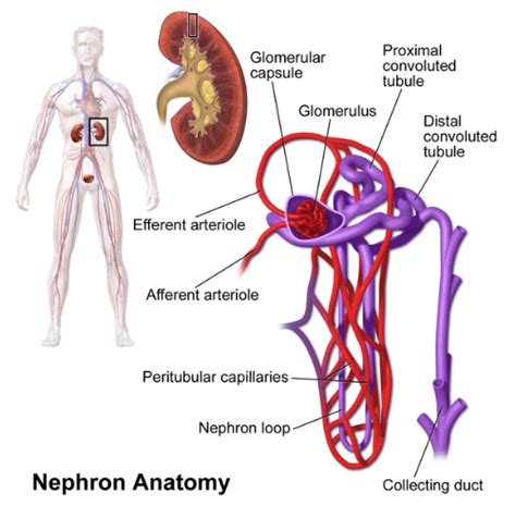
Table of Contents
What is the Structural and Functional Unit of the Kidney?
The kidney, a vital organ in the urinary system, plays a crucial role in maintaining overall body homeostasis. Its primary functions include filtering blood, removing waste products, regulating fluid balance, and producing hormones essential for blood pressure regulation and red blood cell production. Understanding the intricate structure of the kidney is key to comprehending its complex functions. At the heart of this intricate system lies the nephron, the fundamental structural and functional unit of the kidney. This article will delve deep into the nephron's structure, its role in urine formation, and the wider implications of its function for overall health.
The Nephron: A Microscopic Marvel
The human kidney contains approximately one million nephrons, each a remarkably efficient filtration unit. These tiny structures are responsible for the meticulous process of filtering blood, reabsorbing essential nutrients, and excreting waste products in the form of urine. The nephron's complex architecture is directly related to its multifaceted functions. It can be broadly divided into two main parts:
1. The Renal Corpuscle: The Filtration Site
The renal corpuscle, also known as the Malpighian body, is the initial segment of the nephron responsible for filtering blood. It consists of two key components:
-
Glomerulus: This is a network of capillaries enclosed within a cup-like structure called Bowman's capsule. The glomerulus's high-pressure environment is crucial for the process of filtration. The fenestrated endothelium of the glomerular capillaries allows for the passage of water and small solutes, while larger molecules like proteins and blood cells are generally prevented from entering the filtrate. The unique structure of the glomerular capillaries, with their specialized endothelial cells and podocytes (discussed below), ensures a highly selective filtration process.
-
Bowman's Capsule (Glomerular Capsule): This double-walled cup surrounds the glomerulus, collecting the filtered fluid—the glomerular filtrate—which then proceeds to the renal tubule. The visceral layer of Bowman's capsule is composed of specialized epithelial cells called podocytes, which play a critical role in regulating filtration by forming filtration slits between their foot processes. These slits act as a final barrier, preventing the passage of larger molecules. The parietal layer forms the outer wall of the capsule.
2. The Renal Tubule: Fine-Tuning the Filtrate
The renal tubule, a long, convoluted tube extending from Bowman's capsule, is the site where the composition of the filtrate is precisely adjusted. It can be further divided into distinct segments:
-
Proximal Convoluted Tubule (PCT): This initial segment of the renal tubule is characterized by its extensive microvilli, creating a large surface area for reabsorption. The PCT is responsible for the bulk reabsorption of essential nutrients like glucose, amino acids, and electrolytes (sodium, potassium, chloride, etc.) back into the bloodstream. Water is also passively reabsorbed along with these solutes. Secretion of certain substances like hydrogen ions and drugs also occurs in this segment.
-
Loop of Henle (Nephron Loop): This U-shaped structure extends from the PCT into the renal medulla. The Loop of Henle plays a vital role in establishing a concentration gradient in the medulla, critical for concentrating urine. The descending limb of the loop is permeable to water but relatively impermeable to solutes, while the ascending limb is impermeable to water but actively transports sodium, potassium, and chloride ions out of the filtrate. This countercurrent multiplier system creates a hypertonic medullary environment, facilitating water reabsorption in the collecting duct.
-
Distal Convoluted Tubule (DCT): The DCT is responsible for fine-tuning the electrolyte balance in the filtrate. It is influenced by hormones such as aldosterone (which increases sodium reabsorption and potassium secretion) and parathyroid hormone (which increases calcium reabsorption). The DCT further adjusts the composition of the filtrate before it enters the collecting duct.
-
Collecting Duct: This segment, shared by multiple nephrons, receives filtrate from the DCTs of several nephrons. The collecting duct plays a crucial role in regulating water reabsorption under the influence of antidiuretic hormone (ADH). ADH increases the permeability of the collecting duct to water, allowing for greater water reabsorption and the production of concentrated urine. The final product, urine, then flows into the renal pelvis and subsequently to the ureter, bladder, and urethra for excretion.
Nephron Types and Their Functional Significance
Not all nephrons are created equal. Two main types of nephrons exist, differentiated by the length of their Loop of Henle and their location within the kidney:
-
Cortical Nephrons: These nephrons constitute the majority (approximately 85%) of the nephrons and have short Loops of Henle that extend only slightly into the medulla. They are primarily involved in filtering blood and reabsorbing essential nutrients.
-
Juxtamedullary Nephrons: These nephrons have long Loops of Henle that extend deep into the medulla. They play a crucial role in the concentration of urine, contributing significantly to the countercurrent multiplier system that establishes the medullary osmotic gradient. The longer Loop of Henle in juxtamedullary nephrons allows for greater water reabsorption and the production of concentrated urine, essential for maintaining fluid balance, particularly in conditions of dehydration.
Juxtaglomerular Apparatus: Regulation of Blood Pressure and Filtration
The juxtaglomerular apparatus (JGA) is a specialized structure located at the junction of the afferent arteriole, efferent arteriole, and distal convoluted tubule. It plays a crucial role in regulating blood pressure and glomerular filtration rate (GFR). The JGA consists of:
-
Juxtaglomerular cells: These specialized cells in the afferent arteriole secrete renin, an enzyme crucial for the renin-angiotensin-aldosterone system (RAAS). The RAAS is a hormonal pathway that regulates blood pressure by constricting blood vessels and increasing sodium reabsorption.
-
Macula densa: These specialized cells in the distal convoluted tubule detect changes in sodium chloride concentration in the filtrate. They communicate with the juxtaglomerular cells to regulate renin release. If sodium chloride concentration is low, the macula densa signals the juxtaglomerular cells to release more renin, leading to an increase in blood pressure.
Clinical Significance of Nephron Function
The health and proper functioning of nephrons are paramount for overall health. Damage to nephrons, whether due to disease, injury, or aging, can lead to a range of debilitating conditions:
-
Chronic Kidney Disease (CKD): This progressive loss of nephron function is often associated with diabetes, hypertension, and other underlying conditions. CKD can lead to a buildup of waste products in the blood, fluid retention, electrolyte imbalances, and ultimately kidney failure.
-
Acute Kidney Injury (AKI): This sudden loss of kidney function can be caused by various factors such as infections, medications, or dehydration. AKI can be reversible in some cases, but severe cases can lead to kidney failure.
-
Glomerulonephritis: This inflammatory condition affects the glomeruli, impairing their ability to filter blood effectively. It can lead to proteinuria (protein in the urine) and hematuria (blood in the urine).
-
Polycystic Kidney Disease (PKD): This genetic disorder leads to the formation of numerous cysts in the kidneys, impairing their function over time.
Understanding the structure and function of the nephron is crucial for diagnosing and managing kidney diseases. Early detection and appropriate management are vital for preserving kidney function and overall health.
Conclusion: The Nephron's Unparalleled Importance
The nephron, the structural and functional unit of the kidney, is a remarkable example of biological engineering. Its complex architecture facilitates the precise filtration, reabsorption, and secretion processes necessary for maintaining fluid balance, electrolyte homeostasis, and the removal of waste products. The interplay between the different segments of the nephron, regulated by hormonal and neural mechanisms, ensures the kidney's ability to adapt to varying physiological demands. Disruptions in nephron function have profound implications for overall health, highlighting the critical importance of maintaining the health of these microscopic marvels. Continued research into nephron biology and pathophysiology is crucial for developing effective strategies for preventing and treating kidney diseases. The more we understand about the intricacies of the nephron, the better equipped we are to safeguard this vital organ and protect overall human health.
Latest Posts
Latest Posts
-
What Organelles Do Plants Have That Animals Dont
Apr 02, 2025
-
Select The Five Major Mechanisms Of Antimicrobial Resistance
Apr 02, 2025
-
What Are The Three Characteristics Of All Metals
Apr 02, 2025
-
Using The General Properties Of Reaction Enthalpy
Apr 02, 2025
-
Graphing Sine And Cosine Worksheet Answers
Apr 02, 2025
Related Post
Thank you for visiting our website which covers about What Is The Structural And Functional Unit Of The Kidney . We hope the information provided has been useful to you. Feel free to contact us if you have any questions or need further assistance. See you next time and don't miss to bookmark.
