Where Are The Cell Bodies For The Sensory Neurons Located
Muz Play
Apr 03, 2025 · 6 min read

Table of Contents
Where Are the Cell Bodies for Sensory Neurons Located? A Comprehensive Guide
The human nervous system is a marvel of biological engineering, responsible for everything from the simplest reflexes to complex cognitive functions. Understanding its intricate structure is crucial to appreciating its capabilities. A key element of this understanding lies in the location of neuronal cell bodies, particularly those belonging to sensory neurons. This article delves deep into the diverse locations of these cell bodies, exploring the various types of sensory neurons and the implications of their anatomical arrangement.
Understanding Sensory Neurons and Their Function
Before diving into the locations of sensory neuron cell bodies, let's establish a foundational understanding of what these neurons are and what they do. Sensory neurons, also known as afferent neurons, are responsible for transmitting sensory information from the periphery of the body to the central nervous system (CNS), which includes the brain and spinal cord. This information encompasses a wide range of sensations, including:
- Touch: Pressure, temperature, pain, vibration
- Sight: Light detection via photoreceptors in the retina
- Hearing: Sound wave detection in the cochlea of the inner ear
- Taste: Chemical detection on the tongue
- Smell: Chemical detection in the nasal cavity
- Proprioception: Sense of body position and movement
- Balance: Detection of equilibrium and spatial orientation
These sensory experiences are initiated by specialized receptor cells located throughout the body. These receptors transduce various stimuli (light, sound, pressure, etc.) into electrical signals that are then transmitted by sensory neurons to the CNS for processing and interpretation.
The Structure of a Sensory Neuron
Sensory neurons share a common structural characteristic: they are pseudounipolar. This means that they have a single axon that branches into two extensions: a peripheral process and a central process.
- Peripheral process: This extends from the receptor to the cell body. It's the part that directly interacts with the sensory stimulus.
- Central process: This extends from the cell body into the CNS. It carries the sensory information towards the brain or spinal cord.
The cell body, or soma, contains the neuron's nucleus and other essential organelles. Its location is critical because it dictates the pathway sensory information takes to reach the brain.
Locations of Sensory Neuron Cell Bodies: A Detailed Breakdown
The location of sensory neuron cell bodies is not uniform across all sensory modalities. They are strategically located to optimize the transmission of sensory information to the CNS. We can broadly categorize them into:
1. Dorsal Root Ganglia (DRG) of the Spinal Cord
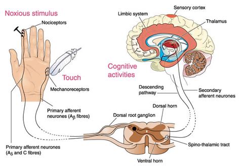
The vast majority of sensory neuron cell bodies associated with somatic sensation (touch, temperature, pain, proprioception) are found in the dorsal root ganglia (DRG). These ganglia are located along the spinal cord, forming clusters of neuronal cell bodies just outside the spinal cord itself. Each DRG serves a specific dermatome – a specific area of skin innervated by a single spinal nerve. The sensory information from the periphery travels through the peripheral process of the sensory neuron to the DRG. The central process then enters the spinal cord via the dorsal root, relaying the information to the brain.
2. Cranial Nerve Ganglia
Sensory information from the head and neck is relayed to the brain via cranial nerves. Unlike spinal nerves, cranial nerves originate directly from the brainstem. The cell bodies for sensory neurons associated with cranial nerves are located in various cranial nerve ganglia. These ganglia are located close to the brainstem and are specific to each cranial nerve involved in sensory function. Examples include:
- Trigeminal ganglion: Houses cell bodies of sensory neurons that innervate the face, scalp, and oral cavity (touch, temperature, pain).
- Vestibulocochlear ganglion: Contains the cell bodies of auditory and vestibular (balance) sensory neurons.
- Geniculate ganglion: Relays taste information from the anterior two-thirds of the tongue.
- Superior and inferior ganglion of the glossopharyngeal nerve: Transmit taste and sensation from the posterior third of the tongue and pharynx.
3. Retina of the Eye
The sensory neurons responsible for vision – the photoreceptor cells (rods and cones) – have their cell bodies located directly within the retina, a layer of neural tissue at the back of the eye. The signal transduction process begins in the photoreceptors, and the information is then relayed through other retinal neurons before reaching the optic nerve and the brain. In this case, the cell bodies are integrated into the sensory organ itself, a unique arrangement compared to other sensory systems.
4. Cochlea of the Inner Ear
Similar to the retina, the cell bodies of sensory neurons responsible for hearing are located within the sensory organ, the cochlea of the inner ear. These are the spiral ganglion neurons, their cell bodies residing within the modiolus, the central axis of the cochlea. The hair cells in the cochlea transduce sound waves into electrical signals, which are then transmitted to the spiral ganglion neurons and relayed to the brain via the vestibulocochlear nerve.
5. Olfactory Epithelium
The sense of smell involves specialized olfactory receptor neurons located in the olfactory epithelium, a tissue lining the nasal cavity. The cell bodies of these olfactory receptor neurons are found within the olfactory epithelium itself. Their axons project directly into the olfactory bulb of the brain, bypassing most traditional relay stations. This direct connection allows for rapid and efficient transmission of olfactory information.
6. Taste Buds
Taste perception depends on specialized taste receptor cells found within taste buds located on the tongue, soft palate, and epiglottis. The sensory neurons responsible for transmitting taste information are found in ganglia associated with the cranial nerves VII (facial), IX (glossopharyngeal), and X (vagus). The exact location of the cell bodies varies depending on which area of the tongue or mouth is involved.
Clinical Significance of Sensory Neuron Cell Body Location
The location of sensory neuron cell bodies has significant clinical implications. Damage or disruption to these ganglia or their pathways can lead to various sensory deficits. For example:
- Herpes zoster (shingles): This viral infection can affect DRG, resulting in painful skin rashes and sensory disturbances in the affected dermatome.
- Peripheral neuropathy: Damage to peripheral nerves can affect sensory neuron function, causing numbness, tingling, pain, and other sensory impairments.
- Acoustic neuroma: A tumor affecting the vestibulocochlear nerve near the vestibulocochlear ganglion can cause hearing loss, tinnitus, and balance problems.
- Bell's palsy: Inflammation of the facial nerve can cause facial paralysis and loss of taste.
Understanding the precise location of sensory neuron cell bodies is essential for accurate diagnosis and treatment of these and other neurological conditions.
Conclusion
The diverse locations of sensory neuron cell bodies reflect the intricate organization and functional specialization of the nervous system. While most somatic sensory neuron cell bodies reside in the DRG, those for special senses are strategically positioned within the sensory organs themselves (retina, cochlea, olfactory epithelium). This anatomical arrangement facilitates efficient transmission of sensory information, providing a foundation for our perception and interaction with the world. Understanding this complex system is crucial not only for basic neuroscience but also for the diagnosis and treatment of various neurological disorders that affect sensory perception. Further research into the development and function of these cell bodies promises to advance our understanding of the nervous system and improve the lives of those affected by sensory disorders.
Latest Posts
Latest Posts
-
How Do You Write Complex Numbers In Standard Form
Apr 04, 2025
-
Shaft Of The Long Bone Is Called
Apr 04, 2025
-
Example Of Stoichiometry In Real Life
Apr 04, 2025
-
Dna Biology And Technology Dna And Rna Structure
Apr 04, 2025
-
Narcotics Act On The Central Nervous System By Producing A
Apr 04, 2025
Related Post
Thank you for visiting our website which covers about Where Are The Cell Bodies For The Sensory Neurons Located . We hope the information provided has been useful to you. Feel free to contact us if you have any questions or need further assistance. See you next time and don't miss to bookmark.
