Which Layer Of The Arterial Wall Is Responsible For Vasoconstriction
Muz Play
Mar 30, 2025 · 5 min read
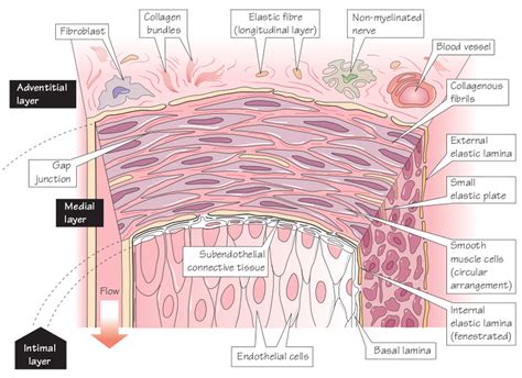
Table of Contents
Which Layer of the Arterial Wall is Responsible for Vasoconstriction?
The regulation of blood pressure and blood flow throughout the body is a complex process, intricately linked to the structure and function of blood vessels. A key component of this regulation is vasoconstriction, the narrowing of blood vessels, which plays a crucial role in maintaining homeostasis. But which layer of the arterial wall is primarily responsible for this vital function? Understanding this requires a detailed examination of the arterial wall's composition and the cellular mechanisms driving vasoconstriction.
The Three Layers of the Arterial Wall
Arteries, the vessels carrying oxygenated blood away from the heart, possess a characteristic three-layered structure:
1. Tunica Intima (Innermost Layer)
The tunica intima is the innermost layer of the arterial wall. It's comprised of:
- Endothelium: A single layer of endothelial cells that form a smooth, continuous lining. These cells are not only crucial for maintaining a non-thrombogenic surface (preventing blood clot formation) but also actively participate in vascular regulation. They release various vasoactive substances, influencing both vasoconstriction and vasodilation.
- Subendothelial Layer: A thin layer of loose connective tissue containing collagen and elastin fibers. This layer provides structural support to the endothelium.
While the endothelium plays a crucial role in vascular tone regulation, it's not the primary layer responsible for vasoconstriction. Its influence is primarily indirect, through the release of signaling molecules that affect the smooth muscle cells in the tunica media.
2. Tunica Media (Middle Layer)
The tunica media is the thickest layer of the arterial wall and is primarily responsible for vasoconstriction. It's composed of:
- Smooth Muscle Cells: These are the key players in vasoconstriction. They are arranged in a circular pattern around the lumen (the central space) of the artery. When these smooth muscle cells contract, the lumen narrows, resulting in vasoconstriction. This contraction is controlled by various neurotransmitters, hormones, and local factors.
- Elastic Fibers and Collagen: These extracellular matrix components provide structural support and elasticity to the tunica media, allowing the artery to withstand the pressure of blood flow and recoil after each heartbeat. The proportion of elastic fibers and smooth muscle cells varies depending on the type of artery (e.g., elastic arteries vs. muscular arteries).
The smooth muscle cells within the tunica media are the primary effectors of vasoconstriction. Their contractile activity is regulated by a complex interplay of factors, as discussed in more detail below.
3. Tunica Externa (Outermost Layer)
The tunica externa, also known as the tunica adventitia, is the outermost layer of the arterial wall. It's composed of:
- Connective Tissue: Primarily composed of collagen and elastin fibers, providing structural support and anchoring the artery to surrounding tissues.
- Nerves and Blood Vessels (Vasa Vasorum): These supply the outer layers of the artery with oxygen and nutrients, as the inner layers are nourished by diffusion from the blood within the lumen.
The tunica externa plays a relatively minor role in vasoconstriction directly. Its primary functions are structural support and nutrient supply.
Mechanisms of Vasoconstriction in the Tunica Media
The smooth muscle cells of the tunica media are innervated by the sympathetic nervous system. Sympathetic nerve fibers release norepinephrine, a potent vasoconstrictor. Norepinephrine binds to α1-adrenergic receptors on the smooth muscle cells, leading to:
- Increased intracellular calcium: This triggers a cascade of events culminating in the contraction of the actin and myosin filaments within the smooth muscle cells.
- Lumen Narrowing: The contraction of the circularly arranged smooth muscle cells causes the lumen of the artery to narrow, reducing blood flow.
Besides sympathetic nervous system activation, several other factors contribute to vasoconstriction:
- Hormones: Angiotensin II, a powerful vasoconstrictor produced by the renin-angiotensin-aldosterone system (RAAS), plays a significant role in blood pressure regulation. Vasopressin (antidiuretic hormone) also acts as a vasoconstrictor, especially during periods of hypovolemia (low blood volume).
- Local Factors: Endothelin-1, a peptide released by endothelial cells, is a potent vasoconstrictor. Other local factors such as prostaglandins and thromboxanes can also influence vascular tone.
- Other Neurotransmitters: Serotonin and dopamine also can influence vasoconstriction, albeit with more complex and nuanced effects compared to norepinephrine.
Clinical Significance of Vasoconstriction and the Tunica Media
Dysregulation of vasoconstriction can have profound clinical consequences, contributing to various cardiovascular diseases. Conditions such as:
- Hypertension (High Blood Pressure): Excessive vasoconstriction is a major contributor to hypertension. The continuous narrowing of blood vessels increases peripheral resistance, elevating blood pressure.
- Raynaud's Phenomenon: This condition involves episodic vasospasm (excessive vasoconstriction) in the arterioles of the extremities, leading to discoloration and pain in the fingers and toes.
- Peripheral Artery Disease (PAD): Atherosclerosis, the buildup of plaque within the arteries, can impair the ability of the tunica media to regulate vasoconstriction and vasodilation, leading to reduced blood flow to the extremities.
- Stroke and Myocardial Infarction: Abnormal vasoconstriction can contribute to the formation of blood clots, which can block blood flow to the brain (stroke) or heart (myocardial infarction).
Conclusion: The Tunica Media's Central Role
In summary, while the endothelium and other layers of the arterial wall contribute indirectly to vascular tone regulation, the tunica media, specifically its smooth muscle cells, is the primary layer responsible for vasoconstriction. Its intricate regulatory mechanisms, involving the sympathetic nervous system, hormones, and local factors, are crucial for maintaining blood pressure and distributing blood flow efficiently throughout the body. Understanding the complexities of this process is crucial for diagnosing and treating various cardiovascular diseases associated with impaired vasoconstriction. Further research continues to unravel the precise interactions between the various cells and signaling pathways involved in this vital physiological process, leading to potential therapeutic targets for diseases affecting the cardiovascular system. Future advancements may focus on understanding how specific genes influence smooth muscle cell contractility and how that relates to the broader picture of cardiovascular health and disease. A deeper understanding of these intricate processes offers hope for improved diagnostic tools and more effective treatments for a wide range of cardiovascular conditions.
Latest Posts
Latest Posts
-
Is An Atom The Smallest Particle
Apr 01, 2025
-
Central Limit Theorem With Means Calculator
Apr 01, 2025
-
What Is The Difference Between Lactic Acid And Alcoholic Fermentation
Apr 01, 2025
-
Which Compound Is Produced During Regeneration
Apr 01, 2025
-
Periodic Trends Atomic Radius Worksheet Answers
Apr 01, 2025
Related Post
Thank you for visiting our website which covers about Which Layer Of The Arterial Wall Is Responsible For Vasoconstriction . We hope the information provided has been useful to you. Feel free to contact us if you have any questions or need further assistance. See you next time and don't miss to bookmark.
