Cartilage Is Separated From Surrounding Tissues By A Fibrous
Muz Play
Apr 01, 2025 · 6 min read
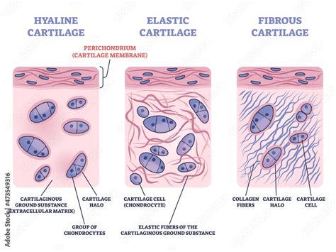
Table of Contents
Cartilage: Separated from Surrounding Tissues by a Fibrous Perichondrium – Structure, Function, and Clinical Significance
Cartilage, a specialized connective tissue, plays a vital role in various bodily functions, providing structural support, facilitating movement, and acting as a shock absorber in joints. A key characteristic of most cartilage is its separation from surrounding tissues by a fibrous perichondrium. Understanding the structure, function, and clinical significance of this perichondrium is crucial to comprehending cartilage biology and its associated pathologies.
The Perichondrium: A Defining Feature of Cartilage
The perichondrium is a dense connective tissue sheath that encloses most cartilage, with the notable exception of articular cartilage (found at the ends of bones in joints) and fibrocartilage (found in intervertebral discs). It's a crucial structure, acting as a boundary, a source of new chondrocytes (cartilage cells), and a provider of nutrients to the avascular cartilage.
Structure of the Perichondrium: The perichondrium is composed of two distinct layers:
-
Outer Fibrous Layer: This layer is primarily composed of dense irregular connective tissue, rich in type I collagen fibers. These fibers provide tensile strength and structural support to the cartilage. It also contains fibroblasts, the cells responsible for producing the collagen fibers. Blood vessels and nerves are abundant in this outer layer, playing a vital role in providing nourishment and innervation to the underlying cartilage, despite the cartilage itself lacking blood vessels.
-
Inner Cellular Layer (Chondrogenic Layer): This layer is characterized by the presence of chondrogenic cells, which are mesenchymal stem cells with the potential to differentiate into chondrocytes. These cells are responsible for the growth and maintenance of the cartilage. They are responsible for appositional growth, the process by which new cartilage is added to the surface of the existing cartilage.
Functional Significance of the Perichondrium
The perichondrium's functions are multifaceted and essential for cartilage health:
-
Nutrient Supply: As cartilage is avascular (lacks blood vessels), the perichondrium serves as a crucial conduit for the diffusion of nutrients, oxygen, and other essential molecules from the surrounding blood vessels to the chondrocytes within the cartilage. This diffusion process is crucial for the survival and function of the cartilage cells.
-
Growth and Repair: The chondrogenic layer of the perichondrium plays a vital role in the growth and repair of cartilage. It contributes to appositional growth, the addition of new cartilage to the surface. While cartilage has limited capacity for self-repair, the perichondrium contributes to this limited regenerative potential. The chondrogenic cells can differentiate into chondrocytes, replenishing the cartilage matrix. However, this repair process is often insufficient for significant cartilage damage.
-
Mechanical Support: The fibrous outer layer provides structural support and protection to the underlying cartilage, protecting it from mechanical stresses and strains. The dense collagen fibers contribute to the overall strength and resilience of the perichondrium and the cartilage it surrounds.
Cartilage Types and Perichondrium
The relationship between cartilage type and the presence or absence of a perichondrium is noteworthy:
-
Hyaline Cartilage: This is the most common type of cartilage, found in the articular surfaces of joints, the nose, trachea, and ribs. Most hyaline cartilage is surrounded by a perichondrium, except for articular cartilage. The perichondrium is crucial for its growth and maintenance.
-
Elastic Cartilage: This type of cartilage is found in the ear and epiglottis. It is also typically surrounded by a perichondrium, contributing to its flexibility and resilience. The elastic fibers within the cartilage, combined with the perichondrium's support, provide the unique properties of this cartilage type.
-
Fibrocartilage: This cartilage is found in intervertebral discs and menisci of the knee. Unlike hyaline and elastic cartilage, fibrocartilage generally lacks a perichondrium. Its structural integrity relies on the dense collagen fiber network within the tissue itself. The lack of a perichondrium is consistent with its limited regenerative capacity.
Clinical Significance of Perichondrial Integrity
The integrity of the perichondrium is critical for cartilage health and repair. Damage or disruption of the perichondrium can significantly impair cartilage function and lead to various pathologies:
-
Articular Cartilage Damage: While articular cartilage lacks a perichondrium, injuries affecting the surrounding tissues can indirectly impact its health. The absence of a perichondrium makes articular cartilage particularly vulnerable to damage and with limited healing capacity.
-
Chondral Defects: These are injuries to the cartilage, often resulting from trauma or overuse. Damage to the perichondrium can hinder the body's natural repair mechanisms, leading to persistent cartilage defects and the development of osteoarthritis.
-
Perichondritis: This is an inflammation of the perichondrium, often caused by infection or trauma. The inflammation can lead to cartilage damage and potentially impair its function. Treatment may involve antibiotics and anti-inflammatory medications.
-
Delayed Cartilage Repair: Damage to the perichondrium can significantly delay or impede cartilage repair after injury. The lack of a complete perichondrium interferes with the delivery of nutrients and the differentiation of chondrogenic cells, hindering the natural regenerative processes.
-
Osteoarthritis: The breakdown of cartilage, a hallmark of osteoarthritis, is often linked to underlying issues with perichondrial integrity. Chronic inflammation, micro-trauma, and genetic factors can contribute to perichondrial damage, exacerbating cartilage degeneration.
Perichondrium and Cartilage Regeneration Strategies
Given the limited self-repair capabilities of cartilage, the perichondrium plays a significant role in developing new regenerative strategies:
-
Perichondrial Grafts: In some surgical procedures, perichondrial grafts are used to promote cartilage repair. These grafts provide a scaffold for the ingrowth of new cartilage cells and support the healing process.
-
Perichondrial Cell-Based Therapies: Research is ongoing to use perichondrial cells in cell-based therapies. These therapies aim to stimulate cartilage regeneration by introducing chondrogenic cells to damaged cartilage.
Conclusion: The Perichondrium – A Critical Player in Cartilage Biology
The fibrous perichondrium is an indispensable component of most cartilage, playing a crucial role in its nutrition, growth, repair, and overall health. Its structure and function are intricately linked to the well-being of the cartilage it surrounds. Understanding the importance of perichondrial integrity is key to comprehending cartilage biology, developing effective diagnostic tools, and designing innovative treatment strategies for cartilage-related pathologies. Further research into the intricacies of the perichondrium and its interaction with the cartilage matrix will undoubtedly lead to advancements in the treatment and prevention of cartilage-related diseases, improving the quality of life for millions affected by these conditions. The ongoing study of the perichondrium's regenerative potential promises to revolutionize cartilage repair strategies in the future.
Future Research Directions
Future research should focus on several key areas:
-
Improving our understanding of the molecular mechanisms that regulate perichondrial cell differentiation and cartilage formation. This will allow for the development of more targeted therapies to stimulate cartilage regeneration.
-
Developing more effective methods for harvesting and culturing perichondrial cells for transplantation. This is crucial for developing successful cell-based therapies.
-
Investigating the role of the perichondrium in the pathogenesis of osteoarthritis and other cartilage-related diseases. This will lead to better preventative and therapeutic strategies.
-
Exploring novel biomaterials and scaffolds that can be combined with perichondrial cells or grafts to enhance cartilage regeneration. This will improve the effectiveness of cartilage repair procedures.
By focusing on these research areas, scientists can pave the way for significant advancements in cartilage repair and treatment, ultimately improving the lives of individuals affected by cartilage-related diseases. The perichondrium remains a key area of focus in the search for effective solutions.
Latest Posts
Latest Posts
-
How To Calculate External Financing Needed
Apr 02, 2025
-
What Is The Bacterial Cell Wall Composed Of
Apr 02, 2025
-
How To Prepare Post Closing Trial Balance
Apr 02, 2025
-
What Would The Potential Of A Standard Hydrogen Electrode
Apr 02, 2025
-
Draw The Tautomer Of This Aldehyde
Apr 02, 2025
Related Post
Thank you for visiting our website which covers about Cartilage Is Separated From Surrounding Tissues By A Fibrous . We hope the information provided has been useful to you. Feel free to contact us if you have any questions or need further assistance. See you next time and don't miss to bookmark.
