Dna Coloring Transcription And Translation Colored
Muz Play
Mar 23, 2025 · 7 min read
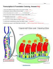
Table of Contents
DNA Coloring, Transcription, and Translation: A Colorful Journey of Genetic Information
The intricate dance of life hinges on the precise transfer of genetic information. This process, encompassing DNA replication, transcription, and translation, is a breathtakingly complex symphony orchestrated by the molecular machinery within our cells. While traditionally depicted in monochrome diagrams, the reality is far more vibrant. Let's delve into the colorful world of DNA, exploring the mechanisms of transcription and translation with a focus on visualizing these crucial processes using color-coding.
The Colorful Canvas of DNA: A Closer Look
DNA, the blueprint of life, is a double helix composed of two intertwined strands. Each strand consists of a chain of nucleotides, each containing one of four nitrogenous bases: adenine (A), guanine (G), cytosine (C), and thymine (T). For our visualization, let's assign each base a distinct color:
- Adenine (A): Green
- Guanine (G): Blue
- Cytosine (C): Red
- Thymine (T): Yellow
This color-coding allows for a readily understandable visual representation of the DNA sequence. Imagine a double helix where one strand displays a sequence like Green-Blue-Red-Yellow (A-G-C-T), and its complementary strand, held together by hydrogen bonds, displays Blue-Green-Yellow-Red (G-C-T-A). This complementary pairing (A with T, and G with C) is crucial for accurate DNA replication and transcription. The very structure itself can be visualized with the phosphate backbone depicted in a contrasting color, perhaps a light grey or even a shimmering silver to highlight its importance in the structural integrity of the DNA molecule.
Transcription: From DNA to RNA – A Colorful Conversion
Transcription is the first step in gene expression, the process by which the information encoded in DNA is used to synthesize proteins. It involves the creation of an RNA molecule that is complementary to a specific DNA sequence. This RNA molecule, messenger RNA (mRNA), carries the genetic code from the DNA in the nucleus to the ribosomes in the cytoplasm, where protein synthesis takes place.
Let's expand our color scheme to include RNA:
- Adenine (A) in RNA: Light Green
- Guanine (G) in RNA: Light Blue
- Cytosine (C) in RNA: Light Red
- Uracil (U) in RNA: Light Yellow
Notice that Uracil (U) replaces Thymine (T) in RNA. During transcription, the enzyme RNA polymerase binds to the DNA at a specific region called the promoter. It then unwinds the DNA double helix, exposing the bases. RNA polymerase uses one DNA strand as a template to synthesize a complementary mRNA molecule.
Imagine a section of DNA with the sequence: Green-Blue-Red-Yellow-Green (A-G-C-T-A). During transcription, the complementary mRNA sequence would be: Light Green-Light Blue-Light Red-Light Yellow-Light Green (U-C-G-A-U). The color change from the deep saturated colors of DNA to the lighter hues of RNA visually distinguishes the two molecules, effectively illustrating the process of transcription. The visualization of the RNA polymerase enzyme itself could be depicted as a small, animated, multi-colored structure moving along the DNA strand, building the mRNA molecule. This animated visual representation further enhances the understanding of the dynamic nature of the transcription process.
Introns and Exons: A Colorful Cut and Paste
Eukaryotic genes are often interrupted by non-coding sequences called introns, interspersed with coding sequences called exons. During RNA processing, introns are removed, and exons are spliced together to form a mature mRNA molecule ready for translation. In our colorful visualization, we could depict introns in a duller, less vibrant shade of their corresponding base colors – a muted Green, Blue, Red, or Yellow, for instance, visually setting them apart from the bright, expressive exons. The splicing process could be animated, showing the introns being excised and the exons joined together, creating a contiguous, vibrant mRNA strand ready for its next journey.
Translation: From RNA to Protein – A Colorful Synthesis
Translation is the second stage of gene expression, where the genetic information encoded in mRNA is used to synthesize a protein. This process takes place in ribosomes, which are complex molecular machines found in the cytoplasm. The mRNA molecule acts as a template for protein synthesis. The genetic code is read in groups of three bases, called codons, each of which specifies a particular amino acid.
We can extend our color scheme to include the 20 standard amino acids. Each amino acid could be assigned a unique color, and we could use a table to show the color-code for each. For example:
- Glycine (Gly): Orange
- Alanine (Ala): Purple
- Valine (Val): Pink
- ...and so on...
During translation, transfer RNA (tRNA) molecules, each carrying a specific amino acid, recognize the codons on the mRNA molecule. The tRNA molecules have anticodons, which are complementary to the codons. The ribosome facilitates the binding of the tRNA molecules to the mRNA molecule and catalyzes the formation of peptide bonds between the amino acids, creating a polypeptide chain.
Imagine a section of mRNA with the sequence: Light Green-Light Blue-Light Red (U-C-G). This codon specifies the amino acid Serine (let's assign it the color Brown). The corresponding tRNA molecule with the anticodon Light Red-Light Blue-Light Green (G-C-A) would deliver a Brown colored Serine to the ribosome. The ribosome would then link this amino acid to the growing polypeptide chain. Each added amino acid would contribute its color to the growing polypeptide chain. The final protein, therefore, would be a vibrant array of colors, each color representing a specific amino acid, visually demonstrating the chain of amino acids creating the final protein product.
Visualizing the Ribosome: A Molecular Machine in Color
The ribosome itself can be visualized as a multi-colored, complex structure. The ribosomal subunits (large and small) could be distinguished by different shades or patterns of colors, highlighting their distinct roles in the translation process. The mRNA passing through the ribosome can be depicted in its light-colored RNA representation while the tRNA molecules, each carrying its unique amino acid color, can be shown attaching and detaching from the mRNA and the growing polypeptide chain. This detailed, color-coded visualization brings to life the dynamic and intricate process of translation.
The Importance of Visualizing Genetic Processes
The use of color-coding in visualizing DNA, transcription, and translation provides several advantages:
- Enhanced Understanding: Color-coding simplifies complex processes by making them visually accessible and easier to comprehend. The distinct colors for each base, RNA molecule, and amino acid make it easier to track the movement and transformation of genetic information.
- Improved Memory Retention: Visual learning aids memory. The color-coded representation of these processes is highly memorable, which helps in retaining the information for longer durations.
- Effective Communication: Color-coding aids in communicating complex scientific concepts to a wider audience, including students and researchers who may not have a deep background in molecular biology.
- Development of Educational Tools: These colorful visualizations can be incorporated into educational materials, such as textbooks, online resources, and interactive simulations, making learning more engaging and effective.
Beyond the Basics: Exploring Advanced Concepts with Color
The color-coding system can also be expanded to visualize more advanced concepts in molecular biology, such as:
- Mutations: Changes in the DNA sequence (e.g., substitutions, insertions, deletions) can be highlighted by changing the color of the affected base or bases. This visual representation instantly shows the impact of mutations on the genetic code.
- Gene Regulation: The binding of regulatory proteins to DNA can be represented by different shapes and colors, illustrating the control mechanisms influencing gene expression.
- Chromatin Structure: The packaging of DNA into chromatin can be depicted using various colors and structures, highlighting the role of histones and other proteins in gene regulation.
The potential applications of color-coding in visualizing genetic processes are vast, providing powerful tools for teaching, research, and communication. By integrating this vibrant and engaging approach into the learning and exploration of molecular biology, we can significantly enhance the understanding of the remarkable processes that underpin life itself. Using color as a means of communication in these complex biological processes opens the door to many new avenues for better visual representation and understanding of the life sciences. The possibilities are limitless, and by employing this exciting methodology, we ensure a clearer and more accessible pathway into the intricate world of molecular biology. The future of teaching and learning within the field will undoubtedly benefit from the enhanced visual aids offered by color-coding techniques.
Latest Posts
Latest Posts
-
Is Boil A Physical Or Chemical Change
Mar 25, 2025
-
How To Write All Real Numbers In Interval Notation
Mar 25, 2025
-
What Are Rows Called On The Periodic Table
Mar 25, 2025
-
What Group Defines Themselves Through A Rejection Of The Mainstream
Mar 25, 2025
-
Transfer Function Of An Rc Circuit
Mar 25, 2025
Related Post
Thank you for visiting our website which covers about Dna Coloring Transcription And Translation Colored . We hope the information provided has been useful to you. Feel free to contact us if you have any questions or need further assistance. See you next time and don't miss to bookmark.
