Draw Cells From The Gram Stained Slide
Muz Play
Mar 16, 2025 · 6 min read
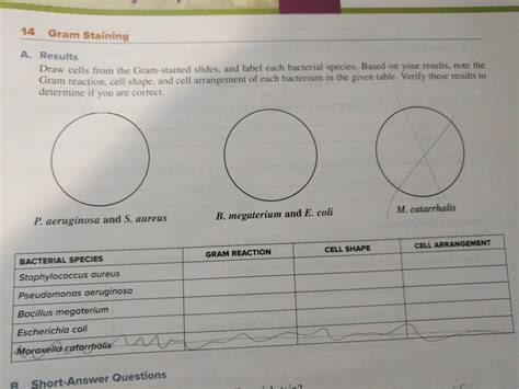
Table of Contents
Drawing Cells from a Gram-Stained Slide: A Comprehensive Guide
Gram staining is a fundamental technique in microbiology, allowing for the differentiation of bacterial cells based on their cell wall composition. This crucial distinction informs diagnosis, treatment strategies, and further microbiological investigation. After performing a Gram stain, accurately drawing the observed cells is a vital skill for any microbiology student or professional. This detailed guide will walk you through the process of creating accurate and informative drawings of Gram-stained bacteria.
Understanding the Importance of Accurate Cell Drawings
Before diving into the specifics of drawing, it's crucial to understand why accurate depictions of Gram-stained cells are essential. These drawings serve several important purposes:
- Record Keeping: Drawings provide a permanent record of the microscopic observations. Photographs can be lost or damaged, but hand-drawn images offer a resilient, readily available record.
- Detailed Observation: The act of drawing forces meticulous observation. You'll notice subtle details – cell morphology, arrangement, size, and staining characteristics – that might be missed during a cursory glance through the microscope.
- Communication: Drawings effectively communicate your findings to others. They are a clear and concise way to share microscopic observations with colleagues, supervisors, or students.
- Improved Understanding: The process of drawing strengthens your understanding of bacterial morphology and the implications of Gram staining.
Materials Needed for Cell Drawing
To create accurate and effective drawings, you'll need a few key materials:
- Microscope: A compound light microscope is essential for observing Gram-stained cells.
- Gram-stained slide: This is your source material. Ensure the slide is properly prepared and has clearly stained bacteria.
- Drawing paper: Use good quality, firm paper that won't bleed or smudge easily. A dedicated microbiology notebook is ideal.
- Pencils: Use a range of pencils – a sharp HB for outlines and finer details, and a softer 2B or 4B for shading and emphasis.
- Eraser: A good quality eraser is crucial for corrections.
- Ruler: A ruler is helpful for accurately estimating cell sizes and maintaining consistent scale.
- Colored pencils (optional): Colored pencils can enhance your drawing by adding color to differentiate Gram-positive and Gram-negative cells (purple and pink/red respectively). However, accuracy of shape and form should remain the priority.
Steps for Drawing Gram-Stained Bacterial Cells
1. Preparing Your Workspace and Microscope
- Clean workspace: Ensure your workspace is clean and well-lit to avoid distractions and smudges.
- Set up your microscope: Prepare your microscope for optimal viewing. Adjust the light intensity and focus carefully. Start with lower magnification to locate the bacterial field and then move to higher magnification for detail.
- Prepare your drawing area: Place your drawing paper comfortably near the microscope, and ensure you have all your drawing materials within easy reach.
2. Observing the Gram-Stained Slide
- Locate the field: Scan the slide systematically under low magnification to locate areas with well-stained and dispersed bacteria. Avoid areas with clumped bacteria or artifacts.
- Increase magnification: Once you've found a suitable field, carefully increase the magnification to achieve optimal detail. Use the fine focus adjustment knob to achieve sharp focus.
- Systematic observation: Begin by observing the overall morphology and arrangement of the bacteria. Note the shape (cocci, bacilli, spirilla), arrangement (chains, clusters, pairs), and size of the cells.
3. Beginning Your Drawing
- Establish a scale: Before you start drawing individual cells, decide on a suitable scale for your drawing. You might choose to draw cells at a 1000x magnification, for instance. Note this scale on your drawing.
- Draw representative cells: Don't attempt to draw every single cell in the field. Select a representative sample of 5-10 cells that accurately reflect the overall morphology and arrangement observed.
- Start with outlines: Begin by drawing the outlines of the cells using a sharp HB pencil. Focus on the accurate representation of shape and size. Use light pencil strokes for initial outlines, allowing for corrections.
4. Adding Details and Shading
- Refine outlines: Once the outlines are complete, refine them by adding details such as cell walls, any visible internal structures (though these may not always be visible with Gram staining), and the overall staining characteristics (purple for Gram-positive and pink/red for Gram-negative).
- Add shading (optional): For added realism, consider adding subtle shading to create depth and texture in your drawing. This can help emphasize the three-dimensional aspect of the cells. Shading should be minimal to avoid obscuring essential morphological details.
- Labeling: Clearly label the drawing with the type of bacteria (if known), the Gram stain result (Gram-positive or Gram-negative), the magnification used, and the date.
5. Reviewing and Refining
- Compare to slide: After completing your drawing, compare it to the cells on the slide under the microscope. Check for accuracy in shape, size, and arrangement.
- Make corrections: Don't be afraid to erase and redraw parts of your drawing to achieve accuracy.
- Final touches: Once you're satisfied with the accuracy and completeness of your drawing, add any final touches, such as a title and any additional notes or observations.
Types of Bacterial Morphologies to Practice Drawing
Practicing drawing different bacterial morphologies will significantly improve your skill and understanding. Here are some common types to focus on:
- Cocci: Spherical or round bacteria. Pay close attention to their arrangement – chains (streptococci), clusters (staphylococci), pairs (diplococci), or tetrads (four cocci in a square).
- Bacilli: Rod-shaped bacteria. Observe variations in length and width. Note if they are arranged in chains (streptobacilli) or palisades (side-by-side).
- Spirilla: Spiral-shaped bacteria. Pay close attention to the number and tightness of the spirals.
- Vibrio: Comma-shaped bacteria.
Advanced Techniques and Considerations
- Scale bars: Incorporate scale bars into your drawings to provide a clear indication of cell size.
- Annotations: Add annotations to highlight specific features of interest, such as unusual morphology or arrangement.
- Digital drawing: If you're comfortable with it, consider using digital drawing software to create your illustrations.
Common Mistakes to Avoid
- Inaccurate size and shape: Ensure that you're accurately representing the size and shape of the cells. Use a ruler to help maintain consistency.
- Over-crowding: Avoid trying to draw too many cells. Focus on representing a representative sample.
- Lack of detail: Include enough detail to accurately portray the morphology and staining characteristics of the bacteria.
- Poor labeling: Always label your drawing clearly with essential information.
Conclusion
Drawing Gram-stained bacterial cells is a valuable skill for any microbiologist. This comprehensive guide provides a step-by-step process for creating accurate and informative drawings. By following these steps and practicing regularly, you can develop your skills and enhance your understanding of bacterial morphology and Gram staining. Remember that accuracy and clarity are key to effective communication and record-keeping in microbiology. Practice makes perfect – the more you draw, the better you'll become at capturing the intricate details of the microbial world. Continuous practice and careful observation are the cornerstones of mastery in this essential skill.
Latest Posts
Latest Posts
-
How To Make A Catalytic Triad
Mar 16, 2025
-
Determine The Reactions At The Supports
Mar 16, 2025
-
A Mixture In Which The Composition Is Uniform Throughout
Mar 16, 2025
-
How To Break The Nitrogen Off An Imine Mechanism
Mar 16, 2025
-
Changes Color At The Endpoint Of A Titration
Mar 16, 2025
Related Post
Thank you for visiting our website which covers about Draw Cells From The Gram Stained Slide . We hope the information provided has been useful to you. Feel free to contact us if you have any questions or need further assistance. See you next time and don't miss to bookmark.
