Identify The Plant Tissues In The Three Images
Muz Play
Mar 28, 2025 · 6 min read
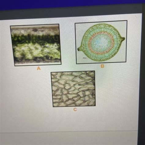
Table of Contents
Identify the Plant Tissues in the Three Images: A Comprehensive Guide
Plant tissues are the fundamental building blocks of plant structures, each with specialized roles in growth, support, and reproduction. Identifying these tissues requires careful observation of cellular structure, arrangement, and overall function. This article will delve into the identification of plant tissues within three hypothetical images (as actual images are not provided). We will explore the key characteristics of each tissue type, allowing you to confidently identify them in various plant samples.
Image 1: A Cross-Section of a Young Stem
This image depicts a cross-section of a young dicot stem. Several distinct tissue types are visible.
1. Epidermis:
- Description: The outermost layer of cells, forming a protective barrier against environmental stressors such as dehydration, pathogens, and physical damage. Cells are typically tightly packed and often covered in a waxy cuticle. In this young stem, the epidermis is a single layer of cells.
- Identification: Look for a single layer of closely packed cells forming the outer boundary. The presence of a cuticle (appearing as a slightly thicker, more transparent layer) is a strong indicator.
- Function: Protection, regulation of water loss, gas exchange (through stomata, if present).
2. Cortex:
- Description: The region between the epidermis and vascular bundles. This image shows a cortex primarily composed of parenchyma cells. Parenchyma cells are relatively large, thin-walled, and irregularly shaped. They are involved in storage, photosynthesis (in some cases), and wound healing. Some sections of the cortex may show the presence of collenchyma cells, providing structural support. These cells have unevenly thickened cell walls.
- Identification: Parenchyma cells appear as large, thin-walled cells with relatively large intercellular spaces. Collenchyma cells are recognizable by their unevenly thickened walls.
- Function: Storage, photosynthesis (in some cases), support (especially collenchyma).
3. Vascular Bundles:
- Description: The vascular bundles are the transport systems of the plant. In dicot stems, these are arranged in a ring. Each bundle contains:
- Xylem: Composed of cells specialized for water and mineral transport. In this young stem, you may see various xylem cell types, including tracheids and vessel elements. Tracheids are long, tapering cells with lignified walls, while vessel elements are shorter and wider, forming continuous tubes.
- Phloem: Responsible for transporting sugars produced during photosynthesis. The phloem is composed of sieve tubes (conducting cells) and companion cells (providing metabolic support to sieve tubes).
- Vascular Cambium: A thin layer of meristematic cells located between the xylem and phloem. This is responsible for secondary growth (increase in stem diameter) in older stems. It is less prominent in this young stem.
- Identification: Xylem cells generally appear darker and thicker-walled than phloem cells. The vascular cambium is a very thin layer of cells between the xylem and phloem.
- Function: Xylem: Water and mineral transport; Phloem: Sugar transport; Vascular Cambium: Secondary growth.
4. Pith:
- Description: The central region of the stem, primarily composed of parenchyma cells. This central core provides storage and support.
- Identification: Similar in appearance to cortical parenchyma but located centrally.
- Function: Storage and support.
Image 2: A Cross-Section of a Leaf
This image shows a cross-section of a typical dicot leaf. The following tissues are readily observable:
1. Upper Epidermis:
- Description: A single layer of closely packed cells forming the upper surface of the leaf. A waxy cuticle is often present, reducing water loss. This epidermis usually lacks stomata (pores for gas exchange).
- Identification: Similar to the stem epidermis but typically without stomata on the upper surface.
- Function: Protection, reducing water loss.
2. Palisade Mesophyll:
- Description: A layer of elongated, columnar cells directly beneath the upper epidermis. These cells contain numerous chloroplasts, the organelles responsible for photosynthesis. They are tightly packed to maximize light absorption.
- Identification: Elongated, columnar cells packed closely together with numerous chloroplasts (which appear as green granules).
- Function: Primary site of photosynthesis.
3. Spongy Mesophyll:
- Description: A layer of irregularly shaped cells beneath the palisade mesophyll. These cells have large intercellular spaces, facilitating gas exchange. They also contain chloroplasts, but fewer than palisade cells.
- Identification: Irregularly shaped cells with large intercellular spaces and fewer chloroplasts than palisade cells.
- Function: Gas exchange, some photosynthesis.
4. Lower Epidermis:
- Description: A single layer of cells forming the lower surface of the leaf. This epidermis contains numerous stomata, surrounded by guard cells that regulate their opening and closing.
- Identification: Similar to the upper epidermis, but containing numerous stomata.
- Function: Gas exchange, regulation of water loss.
5. Vascular Bundles (Veins):
- Description: These are the leaf's veins, composed of xylem and phloem tissues, providing transport pathways for water, minerals, and sugars. They are often embedded within the mesophyll.
- Identification: Similar to vascular bundles in the stem, but often smaller and more extensively branched within the leaf.
- Function: Water and nutrient transport.
Image 3: A Cross-Section of a Root
This image displays a cross-section of a dicot root.
1. Root Cap:
- Description: A protective layer of cells covering the root tip, protecting the delicate meristematic tissues from abrasion as the root grows through the soil. Cells are constantly being replaced.
- Identification: A layer of cells at the very tip of the root, often appearing slightly irregular and loosely packed.
- Function: Protection of the root apical meristem.
2. Root Apical Meristem:
- Description: A region of actively dividing cells located just behind the root cap. These cells give rise to all other root tissues.
- Identification: Smaller, densely packed cells undergoing active cell division. Often located immediately behind the root cap.
- Function: Growth of the root.
3. Zone of Elongation:
- Description: The region where newly formed cells from the apical meristem undergo rapid elongation, contributing to root growth.
- Identification: A region of cells that are increasing in length.
- Function: Root elongation.
4. Zone of Maturation:
- Description: The region where cells differentiate into specialized tissues. This includes the epidermis, cortex, endodermis, pericycle, and vascular cylinder.
- Identification: Cells are differentiating into distinct tissue types.
- Function: Development of mature root tissues.
5. Epidermis (Root Hairs):
- Description: The outermost layer of cells. Root hairs are extensions of epidermal cells, significantly increasing the surface area for water and nutrient absorption.
- Identification: Single layer of cells with numerous root hair extensions.
- Function: Water and nutrient absorption.
6. Cortex:
- Description: A region of parenchyma cells between the epidermis and vascular cylinder. They store food and water.
- Identification: Similar to the cortical parenchyma in the stem.
- Function: Storage, radial transport of water.
7. Endodermis:
- Description: A single layer of cells surrounding the vascular cylinder. Cells are characterized by the Casparian strip, a band of suberin that regulates water movement into the vascular cylinder.
- Identification: A distinct single layer of cells with a visible Casparian strip (often appearing as a slightly darker band).
- Function: Regulation of water and nutrient movement into the vascular cylinder.
8. Pericycle:
- Description: A layer of cells surrounding the vascular cylinder. It's involved in lateral root formation.
- Identification: A layer of cells between the endodermis and vascular cylinder.
- Function: Lateral root formation.
9. Vascular Cylinder (Stele):
- Description: The central region of the root containing the xylem and phloem tissues. In dicot roots, the xylem is arranged in a star-like pattern, with the phloem located between the xylem arms.
- Identification: Central region containing xylem and phloem in a characteristic arrangement.
- Function: Transport of water, minerals, and sugars.
This comprehensive guide provides a detailed description of the key plant tissues found in typical young dicot stems, leaves, and roots. By carefully observing cellular structures, arrangement, and overall function, you can effectively identify these tissues in various plant samples. Remember that variations exist between species, but the general principles of tissue organization and function remain consistent. Further study using microscopic techniques will significantly enhance your ability to accurately identify plant tissues.
Latest Posts
Latest Posts
-
Equations For Motion With Constant Acceleration
Mar 31, 2025
-
Double Replacement Reaction Examples In Real Life
Mar 31, 2025
-
Body Cavities And Membranes Concept Map
Mar 31, 2025
-
What Is Required For Osmosis To Occur
Mar 31, 2025
-
Does Ccl4 Have A Dipole Moment
Mar 31, 2025
Related Post
Thank you for visiting our website which covers about Identify The Plant Tissues In The Three Images . We hope the information provided has been useful to you. Feel free to contact us if you have any questions or need further assistance. See you next time and don't miss to bookmark.
