Image Of Plant And Animal Cell
Muz Play
Mar 28, 2025 · 6 min read
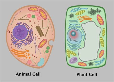
Table of Contents
Delving Deep into the Microscopic World: A Comprehensive Comparison of Plant and Animal Cells
The foundation of all living organisms, from the smallest bacteria to the largest whales, lies within the intricate workings of the cell. While vastly diverse in form and function, all cells share fundamental similarities, primarily their composition and organization. However, significant differences exist between the cellular structures of plants and animals, reflecting their distinct lifestyles and ecological roles. This detailed exploration dives into the fascinating world of plant and animal cells, comparing and contrasting their key features through high-quality imagery and descriptive text.
The Universal Building Blocks: Similarities Between Plant and Animal Cells
Before highlighting the differences, it's crucial to acknowledge the common ground. Both plant and animal cells are eukaryotic cells, meaning they possess a membrane-bound nucleus containing the genetic material (DNA) and other membrane-bound organelles. This sophisticated internal organization distinguishes them from prokaryotic cells, like bacteria, which lack these features. Key shared structures include:
1. Cell Membrane (Plasma Membrane):
The gatekeeper of the cell, the cell membrane is a selectively permeable barrier that regulates the passage of substances in and out. It's primarily composed of a phospholipid bilayer, embedded with proteins and cholesterol. This structure allows for controlled transport of nutrients, waste products, and signaling molecules. (Include a high-quality microscopic image of a cell membrane). The fluidity of this membrane is crucial for cell function.
2. Cytoplasm:
The gel-like substance filling the cell, the cytoplasm houses the organelles and is the site of many metabolic processes. It's primarily composed of water, salts, and various organic molecules. The cytoplasm facilitates intracellular transport and provides a medium for biochemical reactions. (Include a high-quality microscopic image highlighting the cytoplasm and some organelles).
3. Nucleus:
The control center of the cell, the nucleus contains the cell's genetic material, DNA, organized into chromosomes. The nucleus is surrounded by a double membrane called the nuclear envelope, which contains nuclear pores that regulate the transport of molecules between the nucleus and the cytoplasm. (Include a high-quality microscopic image of a nucleus showing the nuclear envelope and nucleolus). The nucleolus, a dense region within the nucleus, is responsible for ribosome production.
4. Ribosomes:
These protein synthesis factories are found in both plant and animal cells. Ribosomes translate the genetic code from messenger RNA (mRNA) into proteins. They can be free-floating in the cytoplasm or attached to the endoplasmic reticulum. (Include a high-quality microscopic image showing ribosomes, possibly attached to the ER).
5. Mitochondria:
Often called the powerhouses of the cell, mitochondria are responsible for cellular respiration, the process of converting glucose into ATP (adenosine triphosphate), the cell's main energy currency. They have their own DNA and are believed to have originated from symbiotic bacteria. (Include a high-quality microscopic image of mitochondria showcasing their characteristic cristae).
6. Endoplasmic Reticulum (ER):
A network of interconnected membranes extending throughout the cytoplasm, the ER plays a crucial role in protein and lipid synthesis. Rough ER, studded with ribosomes, is involved in protein synthesis and modification, while smooth ER is involved in lipid synthesis and detoxification. (Include a high-quality microscopic image showing both rough and smooth ER).
7. Golgi Apparatus (Golgi Body):
The Golgi apparatus is a stack of flattened membrane-bound sacs that processes, packages, and distributes proteins and lipids synthesized by the ER. It modifies and sorts molecules for secretion or transport to other organelles. (Include a high-quality microscopic image of a Golgi apparatus showing its characteristic stacked structure).
8. Lysosomes (in animal cells; analogous structures in plants):
In animal cells, lysosomes are membrane-bound sacs containing digestive enzymes that break down waste materials, cellular debris, and pathogens. Plant cells have vacuoles that perform similar functions, though they have additional roles. (Include a high-quality microscopic image of lysosomes, if possible, or explain the vacuole's analogous function with an image).
Distinguishing Features: Where Plant and Animal Cells Diverge
While sharing these fundamental components, plant and animal cells exhibit significant differences that reflect their distinct roles in nature.
1. Cell Wall: The Defining Feature of Plant Cells
The most striking difference lies in the presence of a rigid cell wall surrounding the plant cell membrane. This wall, primarily composed of cellulose, provides structural support and protection, allowing plants to stand upright and withstand environmental stresses. Animal cells lack this rigid outer layer. (Include a high-quality microscopic image of a plant cell wall, clearly showing its structure in relation to the cell membrane). The cell wall's porosity allows for the passage of water and other small molecules.
2. Chloroplasts: The Photosynthetic Powerhouses
Plant cells contain chloroplasts, organelles responsible for photosynthesis, the process of converting light energy into chemical energy in the form of glucose. This process is vital for plant growth and survival. Chloroplasts contain chlorophyll, the green pigment that absorbs light energy. Animal cells lack chloroplasts and obtain energy by consuming organic matter. (Include a high-quality microscopic image of chloroplasts, showcasing their internal structure and the presence of chlorophyll).
3. Vacuoles: Storage and Support in Plant Cells
Plant cells typically possess a large central vacuole, a membrane-bound sac that occupies a significant portion of the cell's volume. The vacuole serves multiple functions, including storage of water, nutrients, and waste products. It also plays a role in maintaining turgor pressure, the pressure exerted by the cell contents against the cell wall, which helps maintain the plant's structure. Animal cells may have smaller vacuoles, but they are not as prominent as in plant cells. (Include a high-quality microscopic image of a large central vacuole in a plant cell).
4. Plasmodesmata: Intercellular Communication
Plant cells are interconnected by plasmodesmata, channels that connect the cytoplasm of adjacent cells, allowing for communication and transport of molecules between cells. This intercellular connectivity is crucial for plant development and coordination. Animal cells have gap junctions that perform a similar function, but their structure differs. (Include a high-quality microscopic image illustrating plasmodesmata connecting plant cells).
Visualizing the Differences: Microscopic Images and Interpretations
(This section should include multiple high-quality microscopic images of both plant and animal cells at different magnifications. Each image should be clearly labeled and accompanied by a concise description highlighting the key features visible in the image. Consider including images showing different types of plant and animal cells to illustrate diversity.) For example:
- Image 1: A cross-section of a plant leaf cell showing the cell wall, chloroplasts, and large central vacuole.
- Image 2: A transmission electron micrograph of an animal muscle cell showing the numerous mitochondria needed for energy production.
- Image 3: A comparison image showing the relative sizes of plant and animal cells.
- Image 4: A detailed image of a plant cell showing plasmodesmata connecting adjacent cells.
- Image 5: An image highlighting the difference in the structure of the cell membrane in plant and animal cells.
Conclusion: A Tale of Two Cells
The microscopic world reveals a stunning diversity of cellular structures. While plant and animal cells share many fundamental characteristics, their unique adaptations reflect their distinct ecological roles. The presence of a cell wall, chloroplasts, and a large central vacuole are defining features of plant cells, enabling them to perform photosynthesis and maintain their structural integrity. Animal cells, lacking these structures, rely on consuming organic matter for energy and employ different mechanisms for structural support and intercellular communication. Understanding these differences is fundamental to appreciating the complexity and beauty of life at its most basic level. Further exploration into specific cell types and their functions can deepen this understanding, revealing even more fascinating details of this microscopic world.
Latest Posts
Latest Posts
-
According To The Rules Of Osmosis A System Will
Mar 31, 2025
-
Where Are Chondrocytes And Osteocytes Located
Mar 31, 2025
-
List The Types Of Persuasive Speeches
Mar 31, 2025
-
The Energy Needed To Start A Chemical Reaction Is Called
Mar 31, 2025
-
Person In Environment In Social Work
Mar 31, 2025
Related Post
Thank you for visiting our website which covers about Image Of Plant And Animal Cell . We hope the information provided has been useful to you. Feel free to contact us if you have any questions or need further assistance. See you next time and don't miss to bookmark.
