Label The Cavities Of The Head
Muz Play
Mar 27, 2025 · 7 min read
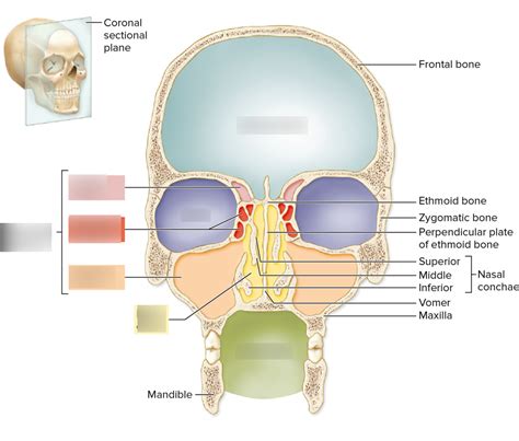
Table of Contents
Labeling the Cavities of the Head: A Comprehensive Guide
The human head, a marvel of biological engineering, houses a complex network of cavities that protect vital organs and facilitate crucial bodily functions. Understanding the anatomy of these cavities is essential for anyone studying medicine, anatomy, or related fields. This comprehensive guide will explore the major cavities of the head, providing detailed descriptions and emphasizing their clinical significance. We'll delve into the boundaries, contents, and clinical correlations of each, ensuring a thorough understanding of this complex region.
Major Cavities of the Head
The head primarily contains two major cavities: the cranial cavity and the facial cavities. These cavities are not completely separate; they are interconnected and share intricate relationships.
1. The Cranial Cavity
The cranial cavity, also known as the intracranial cavity, is the largest cavity within the head. It's a bony enclosure formed by the cranial bones – the frontal, parietal, temporal, occipital, sphenoid, and ethmoid bones. Its primary function is to protect the brain and its associated structures.
Contents of the Cranial Cavity:
- Brain: This is the most significant occupant, consisting of the cerebrum, cerebellum, and brainstem. The intricate folds and convolutions of the brain maximize surface area within the confined space.
- Meninges: Three protective membranes, the dura mater, arachnoid mater, and pia mater, surround the brain, providing cushioning and support. The spaces between these membranes, particularly the subarachnoid space, are crucial for cerebrospinal fluid circulation.
- Cerebrospinal Fluid (CSF): This clear, colorless fluid acts as a shock absorber, protecting the brain from impact. It also provides nutrients and removes waste products.
- Blood Vessels: A vast network of arteries and veins supplies blood to the brain, ensuring a constant supply of oxygen and nutrients. Venous drainage is equally important to remove metabolic waste.
- Cranial Nerves: Twelve pairs of cranial nerves exit the cranial cavity through various foramina (openings) in the cranial bones, connecting the brain to sensory organs and muscles in the head and neck.
Clinical Significance of the Cranial Cavity:
Understanding the cranial cavity's anatomy is critical in diagnosing and treating various neurological conditions. For instance:
- Traumatic Brain Injury (TBI): Impacts to the head can cause fractures of the cranial bones, leading to intracranial hemorrhage (bleeding within the cranial cavity) or brain contusions (bruising).
- Hydrocephalus: An accumulation of excess CSF within the cranial cavity can cause increased intracranial pressure, leading to neurological damage.
- Brain Tumors: Tumors within the cranial cavity can compress brain tissue, causing a range of neurological symptoms depending on the tumor's location and size.
- Meningitis: Infection of the meninges can cause inflammation and swelling, increasing intracranial pressure and potentially leading to severe neurological complications.
- Craniostenosis: Premature fusion of the cranial sutures (joints between the cranial bones) can restrict brain growth, leading to deformities of the skull and potential neurological impairments.
2. The Facial Cavities
The facial cavities are a collection of spaces within the face that contribute to essential functions such as respiration, digestion, and sensory perception. These cavities are interconnected and are often described in relation to their bony boundaries and their specific functions.
a) The Orbital Cavities (Orbits):
The orbits are bony sockets located bilaterally within the skull that house the eyeballs and their associated structures. These cavities are formed by contributions from the frontal, zygomatic, maxillary, ethmoid, sphenoid, and palatine bones.
Contents of the Orbital Cavities:
- Eyeballs: The organs of sight, containing the lens, retina, and other structures necessary for vision.
- Extraocular Muscles: Six muscles control eye movement, allowing for precise gaze control.
- Optic Nerve: This nerve transmits visual information from the retina to the brain.
- Blood Vessels and Nerves: These provide oxygen, nutrients, and innervation to the eye and its associated structures.
- Lacrimal Gland: This gland produces tears, which lubricate and protect the eye.
Clinical Significance of the Orbital Cavities:
Conditions affecting the orbits can severely impair vision and facial function. Some important considerations are:
- Orbital Fractures: Trauma to the face can fracture the orbital bones, leading to displacement of the eyeball (enophthalmos or proptosis) or damage to the optic nerve.
- Orbital Cellulitis: Infection of the tissues surrounding the eye can cause severe swelling and inflammation, potentially leading to vision loss or spread of infection to the brain.
- Thyroid Eye Disease (Graves' Ophthalmopathy): This autoimmune disease can cause bulging of the eyes (exophthalmos) due to inflammation and accumulation of tissue behind the eyes.
b) The Nasal Cavity:
The nasal cavity is a large, air-filled space located within the face, superior to the oral cavity. It's bounded by the nasal bones, maxillae, ethmoid, and sphenoid bones. The nasal cavity plays a crucial role in respiration, filtering, warming, and humidifying inhaled air. It's also involved in olfaction (smell).
Contents of the Nasal Cavity:
- Nasal Septum: A cartilaginous and bony partition dividing the nasal cavity into two halves.
- Nasal Conchae (Turbinates): Bony projections that increase the surface area of the nasal cavity, enhancing air conditioning.
- Nasal Mucosa: A mucous membrane lining the nasal cavity, which contains blood vessels and specialized cells for warming, humidifying, and filtering air. Also contains olfactory receptors for smell.
- Paranasal Sinuses: Air-filled spaces within the bones surrounding the nasal cavity (frontal, maxillary, ethmoid, and sphenoid sinuses). These sinuses contribute to resonance during speech and reduce the weight of the skull.
Clinical Significance of the Nasal Cavity:
Problems in the nasal cavity can range from mild discomfort to severe health issues. Examples include:
- Nasal Fractures: Injuries to the nose can fracture the nasal bones, leading to nasal deformity and breathing difficulties.
- Rhinitis: Inflammation of the nasal mucosa can cause nasal congestion, sneezing, and runny nose. Allergic rhinitis is a common form of rhinitis.
- Sinusitis: Inflammation or infection of the paranasal sinuses can cause facial pain, pressure, and congestion.
- Nasal Polyps: Benign growths in the nasal cavity can obstruct airflow and lead to nasal congestion.
- Nasal Tumors: Though less common, tumors can develop in the nasal cavity or paranasal sinuses, potentially causing obstruction or neurological symptoms.
c) The Oral Cavity (Mouth):
The oral cavity is the entryway to the digestive system, involved in mastication (chewing), taste, and speech. It's bounded by the lips, cheeks, hard and soft palates, and tongue.
Contents of the Oral Cavity:
- Teeth: Structures used for mastication and speech.
- Tongue: A muscular organ involved in taste, speech, and swallowing.
- Salivary Glands: Produce saliva, which aids in digestion and lubrication.
- Oropharynx: The posterior part of the oral cavity that connects to the pharynx.
Clinical Significance of the Oral Cavity:
Conditions impacting the oral cavity can significantly affect overall health. This is because the mouth can be a source and entry point for infections, and issues there can impact eating, speaking and more:
- Dental Caries (Tooth Decay): Bacterial breakdown of tooth enamel leads to cavities.
- Gingivitis and Periodontitis: Inflammation and infection of the gums.
- Oral Cancer: Cancers can develop in the tissues of the oral cavity.
- Oral Thrush: A fungal infection of the mouth.
d) The Middle Ear Cavity (Tympanic Cavity):
The middle ear cavity, located within the temporal bone, plays a critical role in hearing. It's connected to the nasopharynx (upper part of the throat) via the Eustachian tube and to the inner ear via the oval and round windows.
Contents of the Middle Ear Cavity:
- Malleus, Incus, and Stapes: Three tiny bones (ossicles) that transmit vibrations from the tympanic membrane (eardrum) to the inner ear.
- Tympanic Membrane (Eardrum): A thin membrane that vibrates in response to sound waves.
- Middle Ear Muscles: Small muscles that help regulate the transmission of sound vibrations.
Clinical Significance of the Middle Ear Cavity:
Several conditions can affect the middle ear, including:
- Otitis Media (Middle Ear Infection): Infection of the middle ear, often occurring in children.
- Otosclerosis: A disease characterized by abnormal bone growth in the middle ear, affecting hearing.
- Tympanic Membrane Perforation: A rupture of the eardrum.
Interconnections and Clinical Correlations
The cavities of the head are intricately interconnected. For example, infections in the nasal cavity can spread to the paranasal sinuses (sinusitis), and infections in the middle ear can spread to the mastoid air cells (mastoiditis). Similarly, trauma to one area of the head can affect other cavities. A fracture of the orbital floor, for example, can damage the maxillary sinus.
Understanding these interconnections is crucial for effective diagnosis and treatment. Clinicians must consider the possibility of spread of infection or injury between adjacent cavities when evaluating patients with head injuries or infections.
Conclusion
The cavities of the head are essential structures that protect vital organs and enable critical functions. This guide provides a detailed overview of the major cavities, their contents, and their clinical significance. Thorough understanding of this complex anatomy is pivotal for anyone involved in the diagnosis and treatment of head and neck conditions. Further study and exploration of specific pathologies associated with these cavities are encouraged for a deeper appreciation of this fascinating area of human anatomy.
Latest Posts
Latest Posts
-
Is Density And Specific Gravity The Same
Mar 30, 2025
-
Which Complex Carbohydrate Contains Only A 1 4 Glycosidic Linkages
Mar 30, 2025
-
Understanding Media And Culture An Introduction To Mass Communication
Mar 30, 2025
-
The Substance That Is Being Dissolved By A Solvent
Mar 30, 2025
-
What Percentage Of Alcohol Is Absorbed Through The Small Intestine
Mar 30, 2025
Related Post
Thank you for visiting our website which covers about Label The Cavities Of The Head . We hope the information provided has been useful to you. Feel free to contact us if you have any questions or need further assistance. See you next time and don't miss to bookmark.
