Label The Microscopic Structure Of A Skeletal Muscle
Muz Play
Mar 31, 2025 · 6 min read
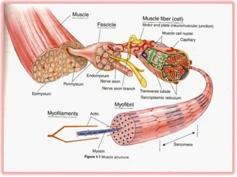
Table of Contents
Labeling the Microscopic Structure of a Skeletal Muscle: A Comprehensive Guide
Understanding the intricate structure of skeletal muscle is crucial for comprehending its function in movement, posture maintenance, and overall bodily health. This detailed guide provides a comprehensive overview of skeletal muscle microscopic anatomy, focusing on the key structural components and their roles. We'll delve into each level of organization, from the whole muscle down to the molecular level, providing clear explanations and illustrative examples to enhance understanding.
Levels of Organization in Skeletal Muscle
Skeletal muscle's impressive strength and precise control stem from its highly organized structure. This organization is hierarchical, with each level building upon the previous one:
1. The Whole Muscle: An Overview
The whole muscle is the macroscopic unit, a distinct organ composed of numerous fascicles bundled together. This structure is surrounded by a tough connective tissue sheath called the epimysium. The epimysium not only provides structural support but also facilitates the transmission of forces generated by the muscle fibers. Consider the biceps brachii, for instance; this is a single muscle comprised of multiple fascicles working in unison.
2. Muscle Fascicles: Bundles of Muscle Fibers
Within the epimysium lie muscle fascicles, bundles of individual muscle fibers. Each fascicle is surrounded by its own connective tissue layer, the perimysium. This perimysium separates individual fascicles, allowing for independent movement and reducing friction between them. The arrangement of fascicles within a muscle varies depending on the muscle's function, influencing its overall strength and range of motion.
3. Muscle Fibers: The Functional Units
The muscle fiber, also known as a muscle cell, is the fundamental unit of skeletal muscle. These cylindrical, elongated cells are multinucleated, meaning they contain multiple nuclei, reflecting their formation through the fusion of myoblasts during development. The muscle fiber is surrounded by the endomysium, a thin layer of connective tissue. The endomysium's delicate nature provides a microenvironment for the muscle fiber while still allowing for efficient nutrient and waste exchange. The sarcolemma, the muscle fiber's cell membrane, plays a vital role in transmitting nerve impulses and regulating the flow of ions, crucial for muscle contraction.
4. Myofibrils: The Contractile Machinery
Within each muscle fiber lie numerous myofibrils, rod-like structures that run the length of the fiber. These myofibrils are the actual contractile elements of the muscle, responsible for generating force. Myofibrils are composed of repeating units called sarcomeres, the basic functional units of muscle contraction.
5. Sarcomeres: The Fundamental Contractile Units
The sarcomere, defined by Z-lines (or Z-discs) at its boundaries, is the smallest unit capable of muscle contraction. This highly organized structure comprises several key proteins:
-
Actin: Thin filaments arranged in a helical pattern, anchored to the Z-lines. Actin filaments contain troponin and tropomyosin, proteins critical for regulating muscle contraction.
-
Myosin: Thick filaments located in the center of the sarcomere, forming the A-band. Myosin molecules have a "head" region that interacts with actin during contraction. The myosin heads possess ATPase activity, hydrolyzing ATP to power the contraction process.
-
Z-lines (or Z-discs): These define the boundaries of the sarcomere and anchor the thin filaments (actin).
-
M-line: Located in the center of the sarcomere, anchoring the thick filaments (myosin).
-
H-zone: The region of the sarcomere containing only thick filaments (myosin), visible only in the relaxed state. This zone shrinks during contraction.
-
A-band (anisotropic band): The entire length of the thick filaments (myosin), including the overlapping regions with thin filaments (actin). This band remains relatively constant in length during contraction.
-
I-band (isotropic band): The region containing only thin filaments (actin), extending from the A-band to the next Z-line. This band shortens during contraction.
The Sliding Filament Theory: A Mechanism for Contraction
The remarkable ability of skeletal muscle to contract and generate force is explained by the sliding filament theory. This theory postulates that muscle contraction results from the sliding of actin filaments over myosin filaments, reducing the distance between Z-lines and shortening the sarcomere. This process requires energy, provided by the hydrolysis of ATP.
The interaction between actin and myosin is regulated by calcium ions (Ca²⁺). When a nerve impulse stimulates a muscle fiber, it triggers the release of Ca²⁺ from the sarcoplasmic reticulum (SR), a specialized intracellular calcium store. The increased Ca²⁺ concentration binds to troponin, causing a conformational change that moves tropomyosin, uncovering the myosin-binding sites on actin. This allows the myosin heads to bind to actin, forming cross-bridges. The power stroke, driven by ATP hydrolysis, pulls the actin filaments toward the center of the sarcomere. The repeated cycling of cross-bridge formation, power stroke, detachment, and reattachment results in the sliding of filaments and muscle contraction.
Specialized Structures within the Muscle Fiber
Beyond the myofibrils, several other structures are essential for muscle fiber function:
-
Sarcoplasmic Reticulum (SR): A network of interconnected tubules surrounding each myofibril. The SR stores and releases Ca²⁺ ions, which are crucial for regulating muscle contraction.
-
Transverse Tubules (T-tubules): Invaginations of the sarcolemma that extend deep into the muscle fiber, allowing for rapid transmission of nerve impulses from the surface of the fiber to the interior. T-tubules ensure that the entire muscle fiber contracts simultaneously.
-
Sarcolemma: The muscle fiber's cell membrane, crucial for the transmission of nerve impulses and the regulation of ion flow.
-
Nuclei: Multiple nuclei are characteristic of skeletal muscle fibers, a result of the fusion of myoblasts during development. These nuclei play a critical role in protein synthesis and overall muscle fiber maintenance.
-
Mitochondria: Abundant mitochondria are present within muscle fibers to provide the ATP necessary for muscle contraction. Their high density reflects the energy demands of muscle activity.
Clinical Significance and Applications
Understanding the microscopic structure of skeletal muscle is not merely an academic exercise; it holds significant clinical implications. Many diseases and conditions directly impact skeletal muscle structure and function. For example:
-
Muscular Dystrophy: This group of genetic disorders affects the structure of muscle proteins, leading to progressive muscle weakness and degeneration. Understanding the underlying structural defects is crucial for developing effective therapies.
-
Myasthenia Gravis: An autoimmune disease characterized by the destruction of acetylcholine receptors at the neuromuscular junction, leading to muscle weakness and fatigue. Knowledge of the neuromuscular junction's structure and function is vital for diagnosis and treatment.
-
Muscle Injuries: Strains, tears, and other muscle injuries often involve damage to muscle fibers, fascicles, or even the whole muscle. Accurate diagnosis and treatment require a thorough understanding of the muscle's hierarchical structure.
-
Muscle Atrophy: The loss of muscle mass and strength, often resulting from disuse, aging, or certain diseases, involves changes at both the microscopic and macroscopic levels. Understanding these changes is important for developing effective preventative and therapeutic strategies.
Conclusion
The microscopic structure of skeletal muscle is a remarkable example of biological organization. The hierarchical arrangement of the whole muscle, fascicles, muscle fibers, myofibrils, and sarcomeres contributes to its exceptional strength, precision, and versatility. A clear understanding of these components, their interactions, and the mechanisms of muscle contraction is fundamental to comprehending both normal muscle function and the pathophysiology of various muscle-related disorders. Continued research into skeletal muscle structure and function promises to unlock further insights into the complexities of human movement and health.
Latest Posts
Latest Posts
-
Effective Nuclear Charge Periodic Table Trend
Apr 01, 2025
-
Examples Of Instantaneous Rate Of Change
Apr 01, 2025
-
How Is The Use Of Symbols Related To Culture
Apr 01, 2025
-
As You Move Across The Periodic Table
Apr 01, 2025
-
What Is In The Atmosphere Of Jupiter
Apr 01, 2025
Related Post
Thank you for visiting our website which covers about Label The Microscopic Structure Of A Skeletal Muscle . We hope the information provided has been useful to you. Feel free to contact us if you have any questions or need further assistance. See you next time and don't miss to bookmark.
