Label The Structures Of The Vertebra
Muz Play
Mar 22, 2025 · 6 min read
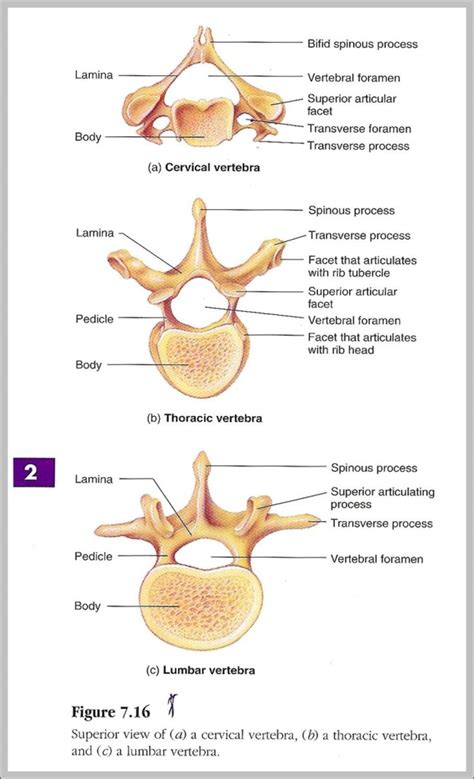
Table of Contents
- Label The Structures Of The Vertebra
- Table of Contents
- Label the Structures of the Vertebra: A Comprehensive Guide
- The Vertebral Column: An Overview
- Anatomy of a Typical Vertebra: A Detailed Breakdown
- 1. Vertebral Body (Corpus Vertebrae):
- 2. Vertebral Arch (Arcus Vertebrae):
- 3. Pedicles (Pedunculi Arcus Vertebrae):
- 4. Laminae (Laminae Arcus Vertebrae):
- 5. Vertebral Foramen (Foramen Vertebrale):
- 6. Spinous Process (Processus Spinosus):
- 7. Transverse Processes (Processus Transversi):
- 8. Superior and Inferior Articular Processes (Processus Articulares Superior et Inferior):
- 9. Intervertebral Foramina:
- Regional Variations in Vertebrae
- Clinical Significance and Applications
- Conclusion
- Latest Posts
- Latest Posts
- Related Post
Label the Structures of the Vertebra: A Comprehensive Guide
Understanding the intricate structure of a vertebra is crucial for anyone studying anatomy, physiology, or related fields. This comprehensive guide will delve into the detailed anatomy of a typical vertebra, exploring each component and its function. We'll equip you with the knowledge to accurately label all the key structures, improving your understanding of the spinal column's complex mechanics.
The Vertebral Column: An Overview
Before we dive into the specifics of a single vertebra, it's important to understand its place within the larger context of the vertebral column, also known as the spine. This column is a flexible yet strong structure composed of 33 vertebrae, divided into five distinct regions:
- Cervical Vertebrae (C1-C7): The seven vertebrae in the neck, characterized by their smaller size and unique features. Atlas (C1) and Axis (C2) are particularly specialized.
- Thoracic Vertebrae (T1-T12): Twelve vertebrae in the chest region, each articulating with the ribs.
- Lumbar Vertebrae (L1-L5): Five large, robust vertebrae in the lower back, bearing the most weight.
- Sacral Vertebrae (S1-S5): Five fused vertebrae forming the sacrum, a triangular bone connecting the spine to the pelvis.
- Coccygeal Vertebrae (Co1-Co4): Four fused vertebrae forming the coccyx (tailbone).
While each region exhibits specific adaptations, a typical vertebra shares many common structural elements. Let's examine these in detail.
Anatomy of a Typical Vertebra: A Detailed Breakdown
A single vertebra, whether cervical, thoracic, or lumbar, consists of several key components:
1. Vertebral Body (Corpus Vertebrae):
- Description: The large, cylindrical anterior portion of the vertebra. It's the weight-bearing part of the vertebra. Its superior and inferior surfaces are covered with hyaline cartilage.
- Function: Supports the weight of the body and provides attachment points for intervertebral discs.
- Clinical Significance: Fractures of the vertebral body are common in trauma, often resulting from compression injuries. Degenerative changes like osteoporosis can also significantly affect the vertebral body.
2. Vertebral Arch (Arcus Vertebrae):
- Description: The posterior part of the vertebra, forming a bony ring around the vertebral foramen. It's composed of the pedicles and laminae.
- Function: Protects the spinal cord and provides attachment points for muscles and ligaments.
- Clinical Significance: Spinal stenosis, a narrowing of the vertebral canal, can compress the spinal cord and nerves. Spondylolysis (a fracture of the pars interarticularis) and spondylolisthesis (forward slippage of one vertebra over another) can occur in the vertebral arch.
3. Pedicles (Pedunculi Arcus Vertebrae):
- Description: Short, thick processes projecting posteriorly from the vertebral body, connecting it to the laminae. They form the sides of the vertebral arch.
- Function: Form the lateral boundaries of the vertebral foramen and provide structural support.
- Clinical Significance: Fractures of the pedicles can lead to instability of the vertebral column.
4. Laminae (Laminae Arcus Vertebrae):
- Description: Flat, broad plates of bone extending posteriorly and medially from the pedicles to meet at the midline, forming the posterior portion of the vertebral arch.
- Function: Complete the bony ring of the vertebral arch and provide attachment points for muscles and ligaments.
- Clinical Significance: Spinal stenosis can be caused by thickening of the laminae. Laminectomy, a surgical procedure involving the removal of portions of the laminae, is sometimes used to relieve pressure on the spinal cord.
5. Vertebral Foramen (Foramen Vertebrale):
- Description: The large opening formed by the vertebral body and vertebral arch, housing the spinal cord.
- Function: Protects the spinal cord and its associated structures.
- Clinical Significance: Conditions such as herniated discs or spinal tumors can impinge on the spinal cord within the vertebral foramen.
6. Spinous Process (Processus Spinosus):
- Description: A single, posterior projection arising from the junction of the laminae. It's palpable along the midline of the back.
- Function: Provides attachment points for muscles and ligaments and serves as a lever for muscle action.
- Clinical Significance: Spinal fractures can involve the spinous process. Palpation of the spinous processes can help in identifying areas of spinal tenderness or abnormalities.
7. Transverse Processes (Processus Transversi):
- Description: Two lateral projections extending from the junction of the pedicles and laminae.
- Function: Provide attachment points for muscles and ligaments, and in the thoracic vertebrae, articulate with the ribs.
- Clinical Significance: Fractures of the transverse processes can occur in trauma.
8. Superior and Inferior Articular Processes (Processus Articulares Superior et Inferior):
- Description: Paired processes projecting superiorly and inferiorly from the junction of the pedicles and laminae. They form the joints between adjacent vertebrae.
- Function: Facilitate movement and provide stability to the vertebral column. They articulate with the inferior articular processes of the vertebra above and the superior articular processes of the vertebra below.
- Clinical Significance: Degenerative joint disease (osteoarthritis) can affect the articular processes, leading to pain and stiffness.
9. Intervertebral Foramina:
- Description: Not part of a single vertebra, but formed by the superior and inferior vertebral notches of adjacent vertebrae. These openings are located laterally.
- Function: Allow passage of spinal nerves from the spinal cord to the rest of the body.
- Clinical Significance: Conditions such as herniated discs or foraminal stenosis can compress spinal nerves, causing pain, numbness, or weakness.
Regional Variations in Vertebrae
While the structures described above represent a typical vertebra, there are significant variations across the different regions of the vertebral column:
-
Cervical Vertebrae: Smaller bodies, transverse foramina for vertebral arteries, bifid spinous processes (except C1 and C7). Atlas (C1) and Axis (C2) are highly specialized, lacking a vertebral body (C1) and possessing the dens (odontoid process) (C2).
-
Thoracic Vertebrae: Heart-shaped bodies, long and sloping spinous processes, costal facets for articulation with ribs.
-
Lumbar Vertebrae: Large, kidney-shaped bodies, thick and short spinous processes, robust transverse processes.
Understanding these regional differences is crucial for accurate identification and interpretation of anatomical structures.
Clinical Significance and Applications
The knowledge of vertebral anatomy is essential in numerous clinical settings:
-
Diagnosis of Spinal Disorders: Radiographic imaging (X-rays, CT scans, MRI) relies heavily on an understanding of vertebral anatomy for accurate interpretation. This helps in diagnosing conditions such as fractures, dislocations, herniated discs, spinal stenosis, and tumors.
-
Surgical Procedures: Spinal surgery, including laminectomy, discectomy, spinal fusion, and instrumentation, requires detailed knowledge of vertebral anatomy to minimize risks and maximize success.
-
Neurological Examination: Understanding the relationship between vertebrae and the spinal cord and nerves is vital for neurological examinations, allowing for accurate localization of neurological deficits.
-
Pain Management: Many musculoskeletal pain conditions originate from the spine. Precise anatomical knowledge helps in pinpointing the source of pain and guiding treatment strategies.
Conclusion
Labeling the structures of the vertebra is more than just an academic exercise; it's the foundation for understanding the complex mechanics and clinical implications of the spinal column. This guide has provided a detailed overview of the anatomy of a typical vertebra and the regional variations. By mastering this knowledge, you’ll gain a significant advantage in any field related to human anatomy, physiology, or medicine. Remember that consistent review and practical application are key to retaining and solidifying this information. Use anatomical models, atlases, and interactive learning tools to reinforce your understanding and achieve accurate labeling of all vertebral structures.
Latest Posts
Latest Posts
-
What Is The Horizontal Rows On The Periodic Table Called
Mar 24, 2025
-
Understanding The Difference Between Strong And Weak Acids
Mar 24, 2025
-
What Are The 3 Parts Of Atp
Mar 24, 2025
-
Does Covalent Compounds Dissolve In Water
Mar 24, 2025
-
What Color Does Magnesium Metal Burn
Mar 24, 2025
Related Post
Thank you for visiting our website which covers about Label The Structures Of The Vertebra . We hope the information provided has been useful to you. Feel free to contact us if you have any questions or need further assistance. See you next time and don't miss to bookmark.
