Osteocytes Sit In Small Chambers Called
Muz Play
Mar 24, 2025 · 7 min read
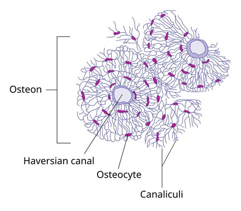
Table of Contents
Osteocytes Sit in Small Chambers Called Lacunae: A Deep Dive into Bone Biology
Osteocytes, the most abundant cells in bone tissue, reside within small spaces called lacunae. Understanding the structure and function of lacunae and their osteocyte inhabitants is crucial to comprehending bone's remarkable properties and its role in overall health. This article delves into the intricate world of osteocytes and lacunae, exploring their development, morphology, interconnectivity, and significance in bone remodeling, mechanotransduction, and systemic health.
The Lacunae: Microscopic Homes for Osteocytes
Lacunae (singular: lacuna) are small, fluid-filled spaces embedded within the bone matrix. These cavities are not simply empty spaces; they are meticulously crafted to house and support osteocytes. The shape of a lacuna often reflects the shape of the osteocyte it contains, with lacunae exhibiting a complex, branching morphology that mirrors the intricate dendritic processes of the osteocytes. These lacunae are not isolated; they are interconnected through a network of microscopic canals.
Composition and Structure of Lacunae
The lacunae are composed of a highly organized extracellular matrix, primarily consisting of collagen fibers and mineralized ground substance. This matrix provides structural support and a controlled microenvironment for the osteocytes. The composition of the matrix surrounding the lacunae can vary depending on the type of bone tissue (e.g., cortical bone versus trabecular bone) and the stage of bone remodeling. The precise chemical composition and organization of this matrix influence the osteocytes' ability to sense mechanical stimuli and communicate with neighboring cells.
Distribution of Lacunae within Bone Tissue
The distribution of lacunae within bone tissue is not random. In cortical bone, lacunae are arranged in a highly organized manner, often aligned along the longitudinal axis of the osteons (Haversian systems). In trabecular bone, the lacunae are more irregularly distributed, reflecting the porous and less organized structure of this bone type. The density of lacunae can vary depending on factors such as age, bone health, and mechanical loading.
Osteocytes: The Master Regulators of Bone
Osteocytes, derived from osteoblasts, are responsible for maintaining bone matrix integrity, sensing mechanical stress, and regulating bone remodeling. Their elongated cell bodies, along with their extensive dendritic processes, occupy the lacunae and extend into the canaliculi. These processes allow for intricate communication with neighboring osteocytes and other bone cells.
The Role of Osteocyte Dendrites
The dendritic processes of osteocytes are vital for communication and nutrient exchange within the bone. These processes extend through tiny canals called canaliculi, forming a vast interconnected network throughout the bone tissue. This network is often referred to as the lacuno-canalicular system. This intricate system facilitates the transport of nutrients, waste products, and signaling molecules between osteocytes and the bone surface. It also plays a key role in mechanotransduction, enabling osteocytes to sense mechanical forces and translate them into biological responses.
Mechanotransduction: Sensing and Responding to Mechanical Stress
Osteocytes are highly sensitive to mechanical stimuli, such as loading and unloading forces. The lacuno-canalicular system plays a crucial role in this process. Fluid flow through the canaliculi, induced by mechanical loading, stimulates osteocytes, initiating signaling pathways that regulate bone formation and resorption. This intricate interplay between mechanical forces and cellular responses ensures that bone adapts to the demands placed upon it. This adaptive response is essential for maintaining bone strength and preventing fractures.
Osteocyte Communication and Bone Remodeling
Osteocytes are not isolated cells. They communicate extensively with each other and with other bone cells, such as osteoblasts and osteoclasts, through various signaling pathways. These pathways involve the release of various factors and ions, modulating bone remodeling processes. Osteocytes act as central regulators of bone remodeling, coordinating the activity of osteoblasts (bone-forming cells) and osteoclasts (bone-resorbing cells) to maintain bone homeostasis. This intricate communication ensures that bone is constantly renewed and adapted to changing mechanical loads.
Osteocyte Dysfunction and Bone Diseases
Dysfunction of osteocytes has been implicated in various bone diseases, including osteoporosis, osteogenesis imperfecta, and other skeletal disorders. Disruptions in the lacuno-canalicular system, impaired osteocyte communication, and altered mechanotransduction can contribute to bone fragility and increased fracture risk. Research continues to explore the role of osteocytes in these diseases, aiming to develop novel therapeutic strategies.
The Lacuno-Canalicular Network: A Highly Organized Communication System
The intricate network of lacunae and canaliculi forms a highly organized communication system within bone tissue. This network facilitates the transport of nutrients, waste products, and signaling molecules between osteocytes and the bone surface. It also plays a key role in mechanotransduction, enabling osteocytes to sense and respond to mechanical stimuli.
Nutrient and Waste Transport
The lacuno-canalicular network provides a pathway for the transport of nutrients and oxygen from blood vessels to osteocytes. Waste products, generated by osteocytes' metabolic activity, are also transported through this network to the bone surface for removal. This continuous exchange of nutrients and waste products is essential for maintaining osteocyte viability and function.
Signaling Molecule Transport
The lacuno-canalicular network also facilitates the transport of signaling molecules between osteocytes and other bone cells. These signaling molecules, including cytokines, growth factors, and other regulatory proteins, play crucial roles in bone remodeling and adaptation to mechanical loads. The efficient transport of these molecules through the network is essential for coordinating bone remodeling activities.
Mechanotransduction within the Network
The fluid flow through the canaliculi, induced by mechanical loading, plays a key role in mechanotransduction. This fluid flow stimulates osteocytes, leading to the production and release of signaling molecules that regulate bone formation and resorption. The lacuno-canalicular network thus acts as a sensor and transducer of mechanical forces, enabling bone to adapt to its mechanical environment.
Clinical Implications: Understanding Osteocytes and Lacunae in Disease
Research on osteocytes and lacunae has significant implications for understanding various bone diseases and developing effective treatments. Alterations in osteocyte function, lacunae morphology, and the lacuno-canalicular network are associated with a range of skeletal disorders.
Osteoporosis
Osteoporosis, a prevalent bone disease characterized by decreased bone mass and increased fracture risk, is often associated with impaired osteocyte function and changes in the lacuno-canalicular network. Research suggests that osteocyte apoptosis (programmed cell death) and reduced osteocyte connectivity contribute to bone fragility in osteoporosis.
Osteogenesis Imperfecta
Osteogenesis imperfecta, also known as brittle bone disease, is a genetic disorder characterized by fragile bones that are prone to fractures. The disease is associated with defects in collagen synthesis, affecting the structural integrity of the bone matrix, including the lacunae and canaliculi. These structural defects impair osteocyte function and communication, further contributing to bone fragility.
Bone Metastasis
Cancer metastasis to bone is a significant clinical problem. Cancer cells can interact with osteocytes, disrupting their normal function and contributing to bone destruction. Understanding the interplay between cancer cells and osteocytes within the lacuno-canalicular network is crucial for developing effective therapies to prevent bone metastasis.
Future Directions and Research
Ongoing research continues to unveil the complexity of osteocytes and their role in bone biology and disease. Advances in imaging techniques, molecular biology, and biomechanics are providing new insights into the function of osteocytes and the lacuno-canalicular network. This knowledge is crucial for developing novel therapeutic strategies for bone diseases and improving bone health overall. Further investigation into specific signaling pathways and the role of the microenvironment surrounding the lacunae are essential to advance our understanding of the complex regulation of bone remodeling. The development of advanced imaging techniques will allow for a more detailed visualization of the lacuno-canalicular network in both health and disease. Ultimately, a greater understanding of these intricate cellular processes will lead to more effective treatments for various bone disorders and improve the overall quality of life for patients affected by these conditions. The field of bone biology is constantly evolving, and future research promises to reveal even more about the vital role of osteocytes and their lacunae homes in maintaining skeletal health.
Latest Posts
Latest Posts
-
In Which Circumstance Would The Courts Find Libel
Mar 27, 2025
-
Solving A Compound Linear Inequality Interval Notation
Mar 27, 2025
-
First Order Versus Zero Order Kinetics
Mar 27, 2025
-
What Are The Vertical Columns Called In The Periodic Table
Mar 27, 2025
-
What Reagent Is Used To Test For Starch
Mar 27, 2025
Related Post
Thank you for visiting our website which covers about Osteocytes Sit In Small Chambers Called . We hope the information provided has been useful to you. Feel free to contact us if you have any questions or need further assistance. See you next time and don't miss to bookmark.
