Parts Of The Compound Light Microscope
Muz Play
Mar 23, 2025 · 6 min read
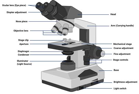
Table of Contents
- Parts Of The Compound Light Microscope
- Table of Contents
- Demystifying the Compound Light Microscope: A Comprehensive Guide to its Parts and Functions
- I. The Optical System: Illuminating the Microscopic World
- A. Light Source (Illuminator): The Foundation of Illumination
- B. Condenser Lens: Focusing the Light
- C. Objective Lenses: Magnifying the Image
- D. Eyepiece (Ocular Lens): The Final Magnification
- II. The Mechanical System: Providing Stability and Control
- A. Microscope Body (Frame): The Sturdy Foundation
- B. Stage: Supporting the Specimen
- C. Focusing Knobs: Achieving Sharp Focus
- D. Revolving Nosepiece (Turret): Selecting the Objective Lens
- III. Essential Accessories: Enhancing Microscopy
- A. Immersion Oil: For High Magnification
- B. Cover Slips: Protecting the Specimen
- C. Slide Preparation Techniques: Critical for Success
- IV. Troubleshooting Common Issues
- V. Conclusion: Mastering the Compound Light Microscope
- Latest Posts
- Latest Posts
- Related Post
Demystifying the Compound Light Microscope: A Comprehensive Guide to its Parts and Functions
The compound light microscope, a cornerstone of biological and medical research, allows us to visualize the intricate details of microscopic life. Understanding its components is crucial for effective use and accurate interpretation of results. This comprehensive guide delves into each part of the compound light microscope, explaining its function and importance in microscopy.
I. The Optical System: Illuminating the Microscopic World
The optical system is the heart of the compound light microscope, responsible for magnifying and illuminating the specimen. It comprises several key components:
A. Light Source (Illuminator): The Foundation of Illumination
Located at the base of the microscope, the light source, or illuminator, provides the illumination necessary to view the specimen. Modern microscopes often use a halogen or LED light source, offering adjustable intensity for optimal viewing. The intensity of the light is crucial; too little light makes the specimen difficult to see, while too much can cause glare and damage the specimen. The light passes through a condenser lens, shaping the light beam before it reaches the specimen.
B. Condenser Lens: Focusing the Light
Positioned beneath the stage, the condenser lens focuses the light from the illuminator onto the specimen. It's adjustable, allowing you to control the cone of light reaching the specimen. A properly adjusted condenser is vital for achieving optimal resolution and contrast. A wider cone of light improves resolution, while a narrower cone enhances contrast, especially with transparent specimens. The condenser typically has an aperture diaphragm, which controls the amount of light passing through it, allowing for fine-tuning of contrast and brightness.
C. Objective Lenses: Magnifying the Image
These lenses are the most important part of the optical system. Located on the revolving nosepiece (turret), a compound microscope usually has multiple objective lenses with different magnification powers (e.g., 4x, 10x, 40x, 100x). The magnification power is engraved on the side of each lens. The 100x objective lens, often referred to as the oil immersion lens, requires immersion oil for optimal resolution and to prevent light scattering. Each objective lens has its own specific working distance (the distance between the lens and the specimen), which must be carefully considered when focusing to prevent damage to the lens or specimen.
D. Eyepiece (Ocular Lens): The Final Magnification
Located at the top of the microscope, the eyepiece (ocular lens) provides the final magnification of the image formed by the objective lens. Typically, eyepieces have a magnification of 10x. The total magnification of the microscope is calculated by multiplying the magnification of the objective lens by the magnification of the eyepiece. For instance, a 40x objective lens combined with a 10x eyepiece provides a total magnification of 400x. Some microscopes have binocular eyepieces, allowing for comfortable viewing with both eyes, reducing eye strain. The interpupillary distance (the distance between the two eyepieces) is adjustable to accommodate individual users.
II. The Mechanical System: Providing Stability and Control
The mechanical system provides the structural support and the mechanisms for focusing and manipulating the specimen. Key components include:
A. Microscope Body (Frame): The Sturdy Foundation
The body or frame of the microscope is the sturdy structure that houses the optical and mechanical components. It provides support and stability, ensuring that the various parts remain aligned and secure during use. The stability of the frame is essential for preventing vibrations that could blur the image. The frame is usually made of metal, contributing to its stability and durability.
B. Stage: Supporting the Specimen
The stage is a flat platform where the specimen slide is placed. It often has stage clips or a mechanical stage to hold the slide in place. A mechanical stage allows for precise movement of the slide using knobs, facilitating easy navigation across the specimen. The stage typically has an aperture in the center to allow light to pass through the specimen.
C. Focusing Knobs: Achieving Sharp Focus
The focusing knobs are used to adjust the distance between the objective lens and the specimen, achieving sharp focus. Most microscopes have two sets of focusing knobs: a coarse adjustment knob for rapid focusing and a fine adjustment knob for precise focusing. Always use the coarse adjustment knob first, then fine-tune the focus with the fine adjustment knob. Incorrect use of these knobs can lead to damage to the specimen or the objective lens.
D. Revolving Nosepiece (Turret): Selecting the Objective Lens
The revolving nosepiece, or turret, is the rotating mechanism that holds the objective lenses. It allows you to easily switch between different objective lenses with different magnification powers. Ensure the objective lens clicks into place securely after rotating the nosepiece. This prevents accidental movement and potential damage to the lenses.
III. Essential Accessories: Enhancing Microscopy
Several accessories can enhance the functionality and capabilities of the compound light microscope:
A. Immersion Oil: For High Magnification
Immersion oil is a special oil with a refractive index similar to glass. It's used with the 100x oil immersion objective lens to improve resolution by minimizing light refraction at the interface between the lens and the slide. Proper application of immersion oil is crucial for achieving optimal resolution at high magnification. Using the oil with other objective lenses is not recommended.
B. Cover Slips: Protecting the Specimen
Cover slips are thin, rectangular pieces of glass placed over the specimen on the slide. They protect the specimen, flatten it, and improve the clarity of the image. Using the appropriate size cover slip is essential for achieving proper focus and image quality. The thickness of the cover slip is also a factor in achieving optimal resolution.
C. Slide Preparation Techniques: Critical for Success
The preparation of microscope slides is a crucial aspect of microscopy. Proper slide preparation ensures that the specimen is properly viewed, and its features are clearly visible. Techniques include staining, fixing, and sectioning, depending on the specimen.
IV. Troubleshooting Common Issues
While the compound light microscope is a robust instrument, certain issues can affect its performance. Understanding and troubleshooting these issues is essential for optimal use.
-
Blurry Image: This could be due to improper focus, dirty lenses, or misaligned components. Clean the lenses with lens paper and ensure that the objective and condenser are properly focused.
-
Insufficient Light: Check the light source intensity and ensure that the condenser is properly aligned and adjusted.
-
Poor Contrast: Adjust the condenser diaphragm or use staining techniques to improve contrast.
-
Oil Immersion Issues: Ensure that proper immersion oil is used and carefully cleaned after use.
V. Conclusion: Mastering the Compound Light Microscope
The compound light microscope is a powerful tool for exploring the microscopic world. Understanding the functions of its various parts, from the light source to the focusing knobs, is paramount for achieving high-quality images and accurate observations. By mastering the instrument and applying proper techniques, users can unlock a world of biological discovery and gain a deeper understanding of the microscopic structures that shape our world. Through diligent practice and consistent care, this sophisticated tool can reveal the intricate beauty of the unseen, transforming curiosity into profound understanding.
Latest Posts
Latest Posts
-
What Is A Subscript In A Chemical Equation
Mar 26, 2025
-
How To Do Post Closing Trial Balance
Mar 26, 2025
-
Como Multiplicar Dos Raices Cuadradas Dividas Entre Otra Raiz Cuadrada
Mar 26, 2025
-
Solid Liquid And Gas Elements In Periodic Table
Mar 26, 2025
-
Integration And Differentiation Of Power Series
Mar 26, 2025
Related Post
Thank you for visiting our website which covers about Parts Of The Compound Light Microscope . We hope the information provided has been useful to you. Feel free to contact us if you have any questions or need further assistance. See you next time and don't miss to bookmark.
