Posterior View Of The Skeletal System
Muz Play
Mar 15, 2025 · 7 min read
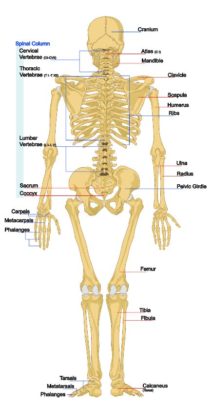
Table of Contents
A Comprehensive Look at the Posterior View of the Skeletal System
The human skeletal system, a marvel of biological engineering, provides the structural framework for our bodies. While often viewed from the anterior (front) perspective, the posterior (back) view reveals a different, equally crucial aspect of its intricate design. This article delves into a detailed exploration of the posterior view of the skeletal system, examining the individual bones, their articulations, and their collective role in supporting, protecting, and enabling movement. We’ll explore key anatomical landmarks, common pathologies, and the importance of understanding this view for healthcare professionals and anatomy enthusiasts alike.
The Vertebral Column: The Backbone of the Posterior View
The vertebral column, or spine, forms the central axis of the posterior skeleton. Its complex structure of 33 vertebrae is divided into five distinct regions:
1. Cervical Vertebrae (C1-C7):
These seven vertebrae in the neck are characterized by their small size and transverse foramina (holes) for the vertebral arteries. Atlas (C1) and Axis (C2) are uniquely shaped to allow for the head's rotation and flexion. The other cervical vertebrae contribute to neck flexibility and support the head's weight. Noteworthy features here include the prominent spinous processes, easily palpable in the neck.
2. Thoracic Vertebrae (T1-T12):
These twelve vertebrae are larger than the cervical vertebrae and articulate with the ribs, forming the thoracic cage. Their unique features include heart-shaped bodies, long downward-sloping spinous processes, and costal facets (articulation points) for rib attachment. This region provides stability and protection for the heart and lungs. Understanding the articulation points between the thoracic vertebrae and ribs is crucial for comprehending respiratory mechanics.
3. Lumbar Vertebrae (L1-L5):
The five lumbar vertebrae are the largest in the vertebral column, reflecting their role in bearing the weight of the upper body. They have robust bodies, short, thick spinous processes, and large vertebral foramina. Their size contributes to their strength and stability, essential for supporting the weight of the torso and transmitting it to the pelvis. Pain in this region is a common complaint, often stemming from issues like herniated discs or spondylolysis.
4. Sacrum:
The sacrum, formed from the fusion of five sacral vertebrae, forms the posterior wall of the pelvis. It articulates superiorly with the lumbar vertebrae (lumbosacral junction) and inferiorly with the coccyx. The sacrum's strong structure is critical for transferring weight from the spine to the hip bones. The sacral foramina, like those in the vertebrae, allow for the passage of nerves.
5. Coccyx:
The coccyx, or tailbone, represents the vestigial remnants of the caudal vertebrae. It's usually composed of three to five fused vertebrae and provides a minor attachment point for muscles and ligaments.
The Rib Cage from a Posterior Perspective
The rib cage, or thoracic cage, viewed posteriorly, shows the twelve pairs of ribs articulating with the thoracic vertebrae. The ribs are categorized into:
- True Ribs (1-7): Directly articulate with the sternum via costal cartilage.
- False Ribs (8-10): Articulate with the sternum indirectly via the costal cartilage of the seventh rib.
- Floating Ribs (11-12): Do not articulate with the sternum.
The posterior view emphasizes the articulation points of the ribs with the thoracic vertebrae, highlighting the importance of the costovertebral and costotransverse joints for respiration and protection of the thoracic organs.
The Pelvic Girdle: Posterior View and its Significance
The pelvic girdle, from a posterior perspective, displays the sacrum, the coccyx, and the two hip bones (ilium, ischium, and pubis). The strong, fused structure of the sacrum and the wide, flaring ilia of the hip bones contribute to the stability and weight-bearing capacity of the pelvis. The sacroiliac joints, where the sacrum articulates with the ilia, are strong and relatively immobile, vital for transferring weight from the spine to the lower limbs. Posterior superior iliac spines (PSIS) and posterior inferior iliac spines (PIIS) are palpable landmarks. Understanding the anatomy of the posterior pelvis is crucial for understanding childbirth, pelvic injuries, and back pain.
The Bones of the Posterior Extremities
The posterior view reveals the bones of the lower limbs, crucial for locomotion and weight-bearing:
- Femur: The thigh bone, the longest and strongest bone in the body. The posterior view shows its prominent linea aspera, a roughened line for muscle attachment.
- Patella: While technically an anterior bone, its articulation with the femur is highly relevant to posterior limb movement and stability.
- Tibia & Fibula: The tibia, or shinbone, is the larger of the two bones in the lower leg. The fibula lies laterally, primarily playing a role in ankle stability. The posterior view clearly shows the tibial tuberosity and the medial and lateral malleoli which form the ankle bones.
- Tarsal, Metatarsal, and Phalangeal Bones: These bones of the foot provide the foundation for balance and weight distribution. The calcaneus, the heel bone, is a significant element visible in the posterior view.
The posterior muscles of the thigh (hamstrings), leg, and foot attach to these bones, making understanding the bony landmarks crucial for comprehending their actions and potential injuries.
The Scapula and Clavicle: Posterior Shoulder Girdle
While much of the scapula (shoulder blade) is seen from the posterior view, the clavicle (collarbone) is primarily anterior. However, the acromion process of the scapula, which articulates with the clavicle, is a significant posterior landmark. This area plays a key role in shoulder movement and stability. The spine of the scapula, a prominent ridge, is also easily palpable in the posterior view.
Clinical Significance of the Posterior Skeletal View
Understanding the posterior view of the skeletal system is critical in various clinical settings:
- Diagnosing Spinal Disorders: The posterior view is essential for assessing spinal curvature (scoliosis, kyphosis, lordosis), identifying vertebral fractures, and evaluating the effects of degenerative conditions like osteoarthritis.
- Assessing Pelvic Injuries: Fractures, dislocations, and other injuries to the sacrum, coccyx, and sacroiliac joints are often diagnosed and treated with careful consideration of the posterior view.
- Musculoskeletal Injuries: Posterior muscle strains and sprains in the back, leg, and shoulder areas are more easily assessed with an understanding of the posterior bone structures and their muscular attachments.
- Neurological Examinations: The posterior view is crucial for assessing the spinal cord and peripheral nerves, as the vertebral column protects the spinal cord and the vertebral foramina allow nerves to exit.
- Surgical Planning: Surgeons rely heavily on a detailed knowledge of the posterior anatomy during spinal surgeries, hip replacements, and other procedures impacting the posterior structures.
Common Pathologies Affecting the Posterior Skeletal System
Many conditions can affect the posterior skeletal system, including:
- Spinal Stenosis: Narrowing of the spinal canal, causing pressure on the spinal cord and nerves.
- Spondylolisthesis: Forward slippage of one vertebra over another.
- Herniated Discs: Protrusion of the intervertebral disc, which can compress nerves.
- Scoliosis: Abnormal lateral curvature of the spine.
- Kyphosis: Excessive outward curvature of the spine (hunchback).
- Lordosis: Excessive inward curvature of the spine (swayback).
- Sacroiliac Joint Dysfunction: Pain and inflammation in the sacroiliac joints.
- Fractures: Bones of the spine, pelvis, and lower limbs are susceptible to fracture.
- Osteoarthritis: Degenerative joint disease affecting the spine and other joints.
Conclusion: The Importance of a Holistic Understanding
The posterior view of the skeletal system, often overlooked, offers a crucial perspective on the human body's structure and function. Its detailed understanding is paramount in healthcare, athletic training, and even artistic representation. By carefully studying the individual bones, their articulations, and their relationships with muscles and ligaments, we gain a deeper appreciation for the complexity and resilience of the human form. This holistic understanding is essential for diagnosing and treating injuries, understanding movement patterns, and appreciating the elegant design of the human skeletal system. The information provided here serves as a foundation for further exploration and deeper study of this vital aspect of human anatomy. Remember always to consult with medical professionals for diagnosis and treatment of any skeletal concerns.
Latest Posts
Latest Posts
-
Difference Between Tlc And Column Chromatography
Mar 15, 2025
-
Energy Required To Remove An Electron From A Gaseous Atom
Mar 15, 2025
-
Que Es La Descomposicion De Acidos
Mar 15, 2025
-
Which Factor Affects Congressional Approval Ratings The Most
Mar 15, 2025
-
Fourier Transform Of A Differential Equation
Mar 15, 2025
Related Post
Thank you for visiting our website which covers about Posterior View Of The Skeletal System . We hope the information provided has been useful to you. Feel free to contact us if you have any questions or need further assistance. See you next time and don't miss to bookmark.
