Simple Columnar Epithelium Under Microscope Labeled
Muz Play
Mar 15, 2025 · 6 min read
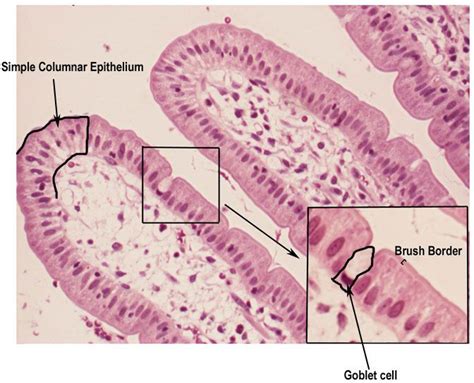
Table of Contents
Simple Columnar Epithelium Under the Microscope: A Comprehensive Guide
Simple columnar epithelium is a type of epithelial tissue characterized by a single layer of tall, column-shaped cells. Understanding its microscopic appearance is crucial for students of histology and those in related medical fields. This comprehensive guide will delve deep into the microscopic features of simple columnar epithelium, including its identification, variations, and clinical significance. We'll explore how to identify it under a microscope, focusing on key features that distinguish it from other epithelial types.
Identifying Simple Columnar Epithelium: Key Microscopic Features
When viewing a prepared slide of simple columnar epithelium under a light microscope, several key characteristics should be readily apparent:
1. Cell Shape and Arrangement:
- Tall and Columnar: The most striking feature is the height of the cells; they are significantly taller than they are wide, resembling columns. This is in stark contrast to squamous epithelium (flat cells) or cuboidal epithelium (cube-shaped cells).
- Single Layer: A crucial distinguishing factor is the presence of only one layer of cells. This is in contrast to stratified columnar epithelium, which has multiple layers. All cells are in direct contact with the basement membrane.
- Nuclei Position: The nuclei of simple columnar epithelial cells are typically located near the base of the cells, appearing elongated and oval-shaped. Their basal location is a helpful distinguishing feature.
2. Cell Borders and Apical Features:
- Lateral Borders: While sometimes indistinct, careful observation may reveal cell borders separating individual columnar cells. Special stains might enhance visualization.
- Apical Surface Specializations: The apical surface, facing the lumen (open space within a tube or organ), may exhibit specialized structures like:
- Microvilli: These are finger-like projections that significantly increase the surface area for absorption. They appear as a brush border under the microscope, often requiring special staining techniques like PAS (Periodic Acid-Schiff) stain for clear visualization. Microvilli are prominent in the simple columnar epithelium lining the small intestine.
- Cilia: These are hair-like projections that move substances along the surface of the epithelium. Cilia appear as fine, hair-like structures extending from the apical surface. They are readily apparent in the simple columnar epithelium of the fallopian tubes. These require careful focusing and possibly specialized stains for optimal viewing.
- Goblet Cells: Often interspersed amongst the columnar cells are goblet cells, specialized cells that secrete mucus. They appear as goblet-shaped cells with a clear, foamy cytoplasm due to the presence of mucus. These are often easily distinguishable with H&E staining.
3. Basement Membrane:
- Delicate Structure: While often subtle, the basement membrane – a thin, supporting layer separating the epithelium from the underlying connective tissue – is present. Special stains (e.g., PAS) are sometimes necessary to highlight its presence.
Variations of Simple Columnar Epithelium
Simple columnar epithelium isn't uniform across the body. Several variations exist, distinguished by their location and the presence of specialized structures.
1. Simple Columnar Epithelium with Microvilli:
This type is predominantly found lining the digestive tract, specifically the small intestine. The presence of abundant microvilli is a defining characteristic, crucial for nutrient absorption. The brush border created by the microvilli is strikingly visible under the microscope, even with standard H&E staining.
2. Simple Columnar Epithelium with Cilia:
This variation is observed in the fallopian tubes (uterine tubes) and parts of the respiratory tract. The cilia's rhythmic beating helps move mucus and other substances along the epithelial surface. The cilia are clearly visible under the microscope as fine, hair-like structures extending from the apical surface.
3. Simple Columnar Epithelium with Goblet Cells:
Goblet cells are frequently found interspersed within simple columnar epithelium lining the digestive and respiratory tracts. Their mucus-secreting function plays a role in lubrication and protection. Their goblet shape and clear cytoplasm readily distinguish them under the microscope.
Distinguishing Simple Columnar Epithelium from Other Epithelial Types
It's crucial to differentiate simple columnar epithelium from other epithelial types. Here's a comparison:
| Feature | Simple Columnar Epithelium | Stratified Columnar Epithelium | Pseudostratified Columnar Epithelium | Simple Cuboidal Epithelium | Simple Squamous Epithelium |
|---|---|---|---|---|---|
| Cell Shape | Tall, columnar | Tall, columnar (multiple layers) | Columnar (appears stratified, but all cells touch basement membrane) | Cube-shaped | Flat |
| Cell Layers | Single | Multiple | Single | Single | Single |
| Nuclei Position | Basal | Varied | Varied | Central | Flattened |
| Apical Features | Microvilli, cilia, goblet cells | May have cilia or goblet cells | Cilia and goblet cells common | None | None |
Clinical Significance of Simple Columnar Epithelium
The health and proper functioning of simple columnar epithelium are vital for various physiological processes. Its disruption can have significant clinical consequences:
- Inflammatory Bowel Disease (IBD): In conditions like Crohn's disease and ulcerative colitis, the simple columnar epithelium lining the digestive tract becomes inflamed and damaged, leading to symptoms like diarrhea, abdominal pain, and weight loss. Microscopic examination of biopsy samples reveals inflammation and changes in the epithelium's structure.
- Gastrointestinal Cancers: Cancers arising from the simple columnar epithelium of the digestive tract, including colorectal cancer, are common. Microscopic examination of cancerous tissue reveals abnormal cell growth and changes in cellular structure and arrangement.
- Respiratory Tract Infections: Damage to the ciliated simple columnar epithelium in the respiratory tract, as seen in infections like bronchitis, impairs the clearance of mucus and debris, leading to increased susceptibility to further infections. Microscopy would show inflammatory cells and potentially damage to the cilia.
- Endometriosis: This condition involves the growth of endometrial tissue outside the uterus. The simple columnar epithelium lining the endometrial cavity can be involved in the disease process. Microscopic analysis of affected tissues is crucial for diagnosis.
- Cervical Cancer: The transformation zone of the cervix, where simple columnar epithelium transitions to stratified squamous epithelium, is a common site for precancerous and cancerous lesions. Microscopic evaluation of Pap smears and biopsies is fundamental for early detection and diagnosis.
Advanced Microscopic Techniques for Studying Simple Columnar Epithelium
Beyond standard light microscopy with H&E staining, various advanced techniques offer enhanced visualization and analysis of simple columnar epithelium:
- Immunohistochemistry (IHC): This technique utilizes antibodies to identify specific proteins within the cells, allowing for detailed analysis of cell types and their function. For example, IHC can identify specific markers for goblet cells or assess the expression of proteins involved in cell signaling and differentiation.
- Electron Microscopy (EM): Both transmission EM (TEM) and scanning EM (SEM) provide ultrastructural detail, allowing for a much closer examination of microvilli, cilia, cell junctions, and other cellular components. TEM reveals intracellular details, while SEM provides three-dimensional views of cell surfaces.
- Confocal Microscopy: This technique allows for the visualization of three-dimensional structures within tissues, providing detailed images of cell layers and specialized structures within the simple columnar epithelium.
Conclusion
Understanding the microscopic features of simple columnar epithelium is essential for accurate diagnosis and treatment of numerous medical conditions. From its characteristic cell shape and arrangement to its diverse apical specializations, the careful observation of this tissue under the microscope is a cornerstone of histopathology and related medical fields. Employing both basic and advanced microscopic techniques provides increasingly detailed insights into its structure, function, and clinical implications. Further exploration into specific variations and the use of advanced microscopic imaging technologies will continue to refine our understanding of this vital epithelial tissue.
Latest Posts
Latest Posts
-
How To Find A Perpendicular Vector
Mar 15, 2025
-
How Could Sulfur Form An Ion
Mar 15, 2025
-
What Elemsnts Are Most Likey To Turn Into Anions Why
Mar 15, 2025
-
What Is The Difference Between Hunger And Appetite
Mar 15, 2025
-
Boiling Point On Graph In Celsius
Mar 15, 2025
Related Post
Thank you for visiting our website which covers about Simple Columnar Epithelium Under Microscope Labeled . We hope the information provided has been useful to you. Feel free to contact us if you have any questions or need further assistance. See you next time and don't miss to bookmark.
