The Cranial Cavity Is A Subdivision Of The
Muz Play
Mar 20, 2025 · 6 min read
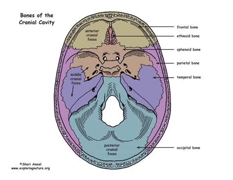
Table of Contents
The Cranial Cavity: A Subdivision of the Dorsal Body Cavity
The human body is a marvel of complex organization, with various cavities housing and protecting vital organs. Understanding the arrangement of these cavities is crucial to comprehending anatomy and physiology. This article delves deep into the cranial cavity, a crucial subdivision of the dorsal body cavity, exploring its structure, contents, and clinical significance.
The Dorsal Body Cavity: A Protective Shell
Before we delve into the specifics of the cranial cavity, it's essential to understand its larger context within the body's overall organizational scheme. The human body possesses two primary cavities: the ventral body cavity and the dorsal body cavity. The ventral cavity, located on the anterior side of the body, houses the thoracic and abdominopelvic cavities, containing organs like the lungs, heart, stomach, and intestines.
In contrast, the dorsal body cavity, situated on the posterior side of the body, is subdivided into two main parts: the cranial cavity and the vertebral canal. The dorsal cavity's primary function is to protect the central nervous system, arguably the most vital control center of the human body.
The Cranial Cavity: A Fortress for the Brain
The cranial cavity, also known as the skull cavity, is the superior portion of the dorsal body cavity. It's a bony enclosure formed by the cranium, a complex structure composed of eight bones intricately joined together by sutures. These sutures, immovable fibrous joints, provide exceptional strength and protection for the brain. The cranial cavity's primary function is to protect the brain from external trauma and to provide a stable environment for its optimal functioning.
The Bones of the Cranial Cavity: A Protective Structure
Let's examine the individual bones that contribute to the cranial cavity's protective shell:
- Frontal Bone: Forms the anterior portion of the skull, contributing to the forehead and the superior portion of the orbits (eye sockets).
- Parietal Bones (2): Situated on either side of the skull, forming the majority of the superior and lateral portions of the cranium.
- Temporal Bones (2): Located on the sides of the skull, inferior to the parietal bones. They house the middle and inner ear structures, along with the mandibular fossa, where the lower jaw articulates.
- Occipital Bone: Forms the posterior portion of the skull, featuring the foramen magnum, a crucial opening through which the spinal cord connects to the brain.
- Sphenoid Bone: A complex, bat-shaped bone located at the base of the skull, contributing to the floor of the cranial cavity and forming part of the orbits and nasal cavity.
- Ethmoid Bone: A delicate bone located anterior to the sphenoid bone, forming part of the nasal septum and contributing to the medial walls of the orbits.
Contents of the Cranial Cavity: More Than Just the Brain
While the brain is the undisputed star resident of the cranial cavity, it's not the only inhabitant. Several other crucial structures call this space home:
-
The Brain: The primary organ of the central nervous system, responsible for controlling virtually all bodily functions, processing information, and enabling higher-level cognitive processes. Its intricate structure, comprising the cerebrum, cerebellum, brainstem, and diencephalon, reflects its complex roles.
-
Meninges: Three protective membranes enveloping the brain: the dura mater, arachnoid mater, and pia mater. These layers provide cushioning, support, and a barrier against infection. The space between the arachnoid and pia mater, the subarachnoid space, contains cerebrospinal fluid (CSF).
-
Cerebrospinal Fluid (CSF): A clear, colorless fluid that circulates within the subarachnoid space and the ventricles of the brain. CSF acts as a cushion, protecting the brain from trauma, transporting nutrients, and removing waste products.
-
Blood Vessels: A complex network of arteries and veins supplies oxygen and nutrients to the brain and removes metabolic waste. The disruption of blood flow to the brain (stroke) can have devastating consequences.
-
Cranial Nerves: Twelve pairs of cranial nerves exit the brain through foramina (openings) in the skull, connecting the brain to various sensory and motor structures in the head and neck. These nerves are crucial for functions like vision, hearing, taste, and facial movement.
Clinical Significance of the Cranial Cavity: A Delicate Balance
The cranial cavity's protective role is paramount for survival. Any compromise to its integrity can lead to severe consequences. Understanding the cranial cavity's clinical significance is crucial for medical professionals:
Head Trauma: A Critical Threat
Traumatic brain injuries (TBIs) are a significant public health concern. These injuries, ranging from mild concussions to severe skull fractures, can result from various mechanisms, including falls, motor vehicle accidents, and sports-related injuries. The severity of a TBI depends on the extent of brain damage and the presence of associated complications such as bleeding (hematoma) or swelling (edema) within the cranial cavity. These complications can increase intracranial pressure, potentially leading to brain herniation—a life-threatening condition.
Craniosynostosis: Premature Closure of Sutures
Craniosynostosis is a congenital condition characterized by the premature fusion of one or more cranial sutures. This premature fusion can lead to abnormal skull shape and potentially affect brain development. Early diagnosis and surgical intervention are often necessary to correct the skull deformity and prevent neurological complications.
Infections: Meningitis and Encephalitis
The meninges and brain are vulnerable to infections. Meningitis, an inflammation of the meninges, and encephalitis, an inflammation of the brain, can be caused by various pathogens, including bacteria, viruses, and fungi. These infections can cause severe neurological symptoms, and prompt treatment is crucial to prevent permanent damage or death.
Tumors: Space-Occupying Lesions
Brain tumors, both benign and malignant, can arise within the cranial cavity. These tumors can exert pressure on surrounding brain tissues, causing neurological deficits depending on their location and size. Treatment options vary depending on the type and location of the tumor and may involve surgery, radiation therapy, or chemotherapy.
Advanced Imaging Techniques: A Window into the Cranial Cavity
Modern medical imaging techniques provide invaluable tools for visualizing the cranial cavity and its contents. These techniques allow for non-invasive assessment of brain structure and function, aiding in the diagnosis and management of various neurological conditions:
-
Computed Tomography (CT) Scans: Generate detailed cross-sectional images of the brain, allowing for the identification of skull fractures, hemorrhages, and other structural abnormalities.
-
Magnetic Resonance Imaging (MRI) Scans: Produce high-resolution images of the brain, providing exceptional visualization of soft tissues, including the brain itself, meninges, and blood vessels. MRI is particularly useful for detecting brain tumors and other subtle abnormalities.
-
Functional MRI (fMRI): Measures brain activity by detecting changes in blood flow. fMRI is used to study brain function in health and disease.
-
Positron Emission Tomography (PET) Scans: Uses radioactive tracers to visualize metabolic activity within the brain. PET scans are useful for detecting brain tumors, assessing the extent of brain damage after stroke, and studying neurological diseases.
Conclusion: The Cranial Cavity—A Complex and Vital Space
The cranial cavity, a crucial subdivision of the dorsal body cavity, serves as a protective fortress for the brain and its associated structures. Its intricate bony architecture, protective membranes, and circulating cerebrospinal fluid work in concert to maintain a stable and optimal environment for brain function. Understanding the anatomy, contents, and clinical significance of the cranial cavity is essential for appreciating the complexity of the human body and the potential consequences of disruptions within this vital space. The advancements in medical imaging have revolutionized our ability to visualize and understand this critical area, leading to improved diagnosis and treatment of neurological conditions. Continued research in this field will undoubtedly lead to further advancements in our understanding and management of diseases affecting the brain and its protective shell.
Latest Posts
Latest Posts
-
Moment Of Inertia Of Rectangular Prism
Mar 20, 2025
-
What Are The Two Starting Materials For A Robinson Annulation
Mar 20, 2025
-
Living Things That Respond To Their Environment
Mar 20, 2025
-
Elements Of Group 17 Are Called
Mar 20, 2025
-
Is A Polymer Of Amino Acids
Mar 20, 2025
Related Post
Thank you for visiting our website which covers about The Cranial Cavity Is A Subdivision Of The . We hope the information provided has been useful to you. Feel free to contact us if you have any questions or need further assistance. See you next time and don't miss to bookmark.
