The Semilunar Valves Prevent Backflow Into The
Muz Play
Mar 30, 2025 · 6 min read
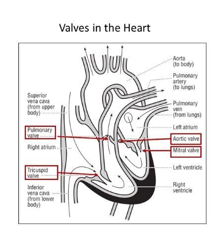
Table of Contents
The Semilunar Valves: Preventing Backflow and Ensuring Efficient Blood Circulation
The human heart, a tireless pump, works tirelessly to circulate blood throughout the body. This intricate process relies heavily on a series of one-way valves that ensure blood flows in the correct direction. Among these crucial valves are the semilunar valves, sentinels guarding against the backflow of blood and maintaining the unidirectional flow essential for life. This article will delve deep into the anatomy, function, and clinical significance of the semilunar valves, exploring how they prevent backflow and contribute to the overall efficiency of the circulatory system.
Understanding the Anatomy of the Semilunar Valves
The heart possesses two sets of semilunar valves: the pulmonary valve and the aortic valve. These valves are strategically positioned at the exits of the heart's ventricles, preventing the retrograde flow of blood.
The Pulmonary Valve: Guarding the Pulmonary Artery
The pulmonary valve is located between the right ventricle and the pulmonary artery. Its primary function is to prevent blood from flowing back from the pulmonary artery into the right ventricle after the right ventricle contracts and ejects blood into the pulmonary circulation. This valve is composed of three cusps, or leaflets, shaped like half-moons (hence the name "semilunar"). These cusps are composed of fibrous connective tissue covered by a thin layer of endocardium, the inner lining of the heart. During ventricular contraction (systole), the pressure within the right ventricle forces the cusps open, allowing blood to flow freely into the pulmonary artery. When the ventricle relaxes (diastole), the pressure in the pulmonary artery exceeds that in the right ventricle, causing the cusps to close, preventing backflow.
The Aortic Valve: Protecting the Aorta
The aortic valve, situated between the left ventricle and the aorta, plays a similar role but on a larger scale. It prevents the backflow of oxygenated blood from the aorta back into the left ventricle after left ventricular contraction. Like the pulmonary valve, it also consists of three semilunar cusps. The left ventricle, being significantly more muscular than the right, generates much higher pressure during contraction, demanding a robust valve to withstand this force. The robust structure of the aortic valve ensures that the oxygen-rich blood propelled into the systemic circulation remains unidirectional, effectively supplying the entire body.
The Mechanics of Semilunar Valve Function: Preventing Backflow
The mechanism by which semilunar valves prevent backflow is a marvel of natural engineering. It involves a precise interplay of pressure gradients, valve cusp anatomy, and ventricular contraction and relaxation cycles.
Pressure Gradients: The Driving Force
The opening and closing of the semilunar valves are primarily driven by pressure differences between the ventricles and their respective arteries. During ventricular systole, the pressure within the ventricles surpasses that in the pulmonary artery and aorta. This pressure difference forces the semilunar cusps open, allowing blood to flow into the arteries. Conversely, during diastole, the ventricular pressure falls below that in the arteries. This pressure reversal causes the cusps to snap shut, effectively sealing off the pathway and preventing backflow.
Cusp Anatomy: Ensuring a Tight Seal
The unique shape and arrangement of the semilunar cusps are crucial for preventing backflow. The cusps are designed to fit snugly together when closed, forming a watertight seal that prevents blood from leaking back into the ventricles. Moreover, the cusps possess small, thickened nodules at their edges called noduli Arantii, which help ensure complete closure and prevent inversion of the cusps under high pressure.
Ventricular Contraction and Relaxation: The Rhythm of Flow
The rhythmic contraction and relaxation of the ventricles dictate the cyclical opening and closing of the semilunar valves. Ventricular contraction creates the necessary pressure to open the valves and propel blood forward. Ventricular relaxation allows the arterial pressure to close the valves and prevent backflow. This coordinated sequence ensures the smooth and efficient flow of blood throughout the circulatory system.
Clinical Significance of Semilunar Valve Disorders
Dysfunction of the semilunar valves can lead to significant cardiovascular complications. These disorders can be congenital (present at birth) or acquired (develop later in life).
Aortic Stenosis: Narrowing of the Aortic Valve
Aortic stenosis refers to the narrowing of the aortic valve orifice, restricting blood flow from the left ventricle into the aorta. This condition can lead to reduced cardiac output, shortness of breath, chest pain, and even heart failure. The causes of aortic stenosis are varied and can include congenital defects, calcification (age-related hardening), or rheumatic heart disease.
Aortic Regurgitation: Backflow Through the Aortic Valve
Aortic regurgitation (or insufficiency) occurs when the aortic valve doesn't close completely during diastole, allowing blood to leak back from the aorta into the left ventricle. This backflow increases the workload on the left ventricle, potentially leading to left ventricular enlargement and heart failure. Causes include congenital abnormalities, connective tissue disorders, and infections.
Pulmonary Stenosis: Narrowing of the Pulmonary Valve
Pulmonary stenosis is the narrowing of the pulmonary valve, restricting blood flow from the right ventricle to the pulmonary artery. This can lead to reduced oxygenation of the blood and right ventricular hypertrophy (enlargement). Congenital defects are the most common cause of pulmonary stenosis.
Pulmonary Regurgitation: Backflow Through the Pulmonary Valve
Pulmonary regurgitation occurs when the pulmonary valve doesn't close properly during diastole, causing blood to leak back from the pulmonary artery into the right ventricle. While often less severe than aortic regurgitation, it can still lead to right ventricular enlargement and heart failure. Congenital defects and pulmonary hypertension are common causes.
Diagnosis and Treatment of Semilunar Valve Disorders
Diagnosing semilunar valve disorders involves a combination of physical examination, electrocardiography (ECG), echocardiography (ultrasound of the heart), and chest X-rays. Echocardiography is particularly crucial, providing detailed images of the valves and assessing their function.
Treatment options vary depending on the severity of the condition and the individual's overall health. Mild cases may only require regular monitoring. More severe cases may necessitate interventions such as:
-
Medication: Medications may be prescribed to manage symptoms, such as diuretics for fluid retention or medications to strengthen the heart muscle.
-
Balloon Valvuloplasty: This minimally invasive procedure uses a balloon catheter to widen a narrowed valve opening.
-
Valve Replacement: In severe cases, surgical replacement of the affected valve may be necessary. This can involve either a mechanical valve or a bioprosthetic valve (made from animal tissue).
Conclusion: The Semilunar Valves - Essential Components of a Healthy Heart
The semilunar valves, the pulmonary and aortic valves, are vital components of the cardiovascular system. Their primary function, preventing backflow of blood, is essential for maintaining efficient and unidirectional blood flow. Understanding their anatomy, function, and the potential for dysfunction is crucial for healthcare professionals and individuals alike. Early diagnosis and appropriate management of semilunar valve disorders are critical in preventing serious cardiovascular complications and improving overall health outcomes. The intricate mechanics of these valves serve as a testament to the remarkable design of the human body and highlight the importance of maintaining cardiovascular health.
Latest Posts
Latest Posts
-
Electric Field Lines About A Point Charge Extend
Apr 01, 2025
-
Does Ionization Energy Increase Across A Period
Apr 01, 2025
-
Integration By Parts How To Choose U And Dv
Apr 01, 2025
-
Protein Synthesis Takes Place In The
Apr 01, 2025
-
Microscopic Anatomy Of A Muscle Fiber
Apr 01, 2025
Related Post
Thank you for visiting our website which covers about The Semilunar Valves Prevent Backflow Into The . We hope the information provided has been useful to you. Feel free to contact us if you have any questions or need further assistance. See you next time and don't miss to bookmark.
