What Color Is A Animal Cell
Muz Play
Apr 02, 2025 · 6 min read

Table of Contents
What Color Is an Animal Cell? The Surprising Answer and Microscopy's Role
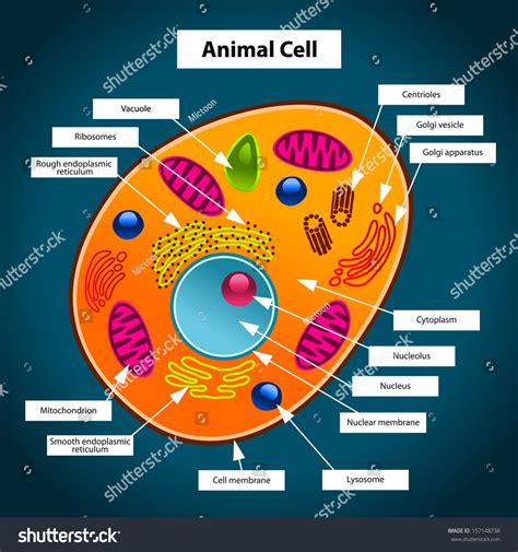
The question, "What color is an animal cell?" might seem simple at first glance. However, the answer is far more nuanced than a single color. Unlike vibrant flowers or colorful insects, animal cells lack the inherent pigmentation that creates readily observable colors. Their appearance under a microscope is primarily dictated by the staining techniques used and the cellular components being observed. This article delves deep into the fascinating world of animal cell visualization, exploring the techniques used to reveal their structures and the reasons behind their seemingly colorless nature.
The Illusion of Colorlessness: Why Animal Cells Appear Clear
Animal cells, unlike plant cells, lack chloroplasts, the organelles responsible for the green color in plants. Chloroplasts contain chlorophyll, a pigment crucial for photosynthesis. The absence of chlorophyll and other prominent pigments contributes significantly to the apparent transparency of animal cells in their natural state. When viewed under a light microscope without staining, they appear as colorless or pale, translucent structures. This lack of inherent color makes them difficult to observe directly without employing various staining and microscopy techniques.
The Power of Staining: Unveiling the Cellular Landscape
Staining techniques are crucial for visualizing the intricate details of animal cells. Different stains target specific cellular components, revealing their structures and functions. The choice of stain dramatically impacts the perceived "color" of the cell. Here are some commonly used stains and their effects:
1. Hematoxylin and Eosin (H&E) Staining: The Gold Standard
H&E staining is a widely used technique in histology, the study of tissues. Hematoxylin, a basic dye, stains acidic components such as DNA and RNA a deep purple or blue. Eosin, an acidic dye, stains basic components such as cytoplasm and extracellular matrix a pinkish-red. Therefore, using H&E staining, cell nuclei appear purplish-blue, while the cytoplasm and other cellular structures appear pinkish-red. The overall appearance of the cell under H&E staining is a blend of these two colors.
2. Wright-Giemsa Stain: Blood Cell Specialist
Wright-Giemsa stain is specifically designed for blood cell staining. This polychromatic stain allows for the differentiation of various blood cell types based on their staining characteristics. It produces a range of colors, including purple, red, and blue, depending on the cellular components and their affinity for the stain. For instance, red blood cells stain a characteristic pinkish-red, while white blood cells exhibit diverse staining patterns reflecting their different cellular components.
3. Periodic Acid-Schiff (PAS) Stain: Carbohydrate Enthusiast
PAS stain is primarily used to detect carbohydrates and glycoproteins. It stains these components a vibrant magenta or pink. This stain is particularly useful in visualizing glycogen deposits in cells and other carbohydrate-rich structures. The overall color of the cell using this stain depends heavily on the concentration of carbohydrates present.
4. Immunohistochemistry (IHC): Targeted Staining for Specific Proteins
IHC is a more advanced technique used to identify and visualize specific proteins within cells. It employs antibodies conjugated to enzymes or fluorescent molecules that bind to target proteins. The color produced depends on the detection method used. For example, chromogenic IHC uses enzyme substrates to produce colored precipitates, enabling visualization of the target protein within the cell. This makes the cells show different colors depending on the protein of interest.
Microscopy Techniques: Illuminating the Cell's Structure
The color of an animal cell as perceived also hinges heavily on the microscopy technique used. Different microscopy methods provide varying levels of detail and contrast, impacting the overall visual impression.
1. Light Microscopy: The Basics
Light microscopy is the most fundamental method. Staining is often crucial to improve contrast and visibility under light microscopy. The color observed depends entirely on the stain used. Without staining, animal cells appear essentially colorless.
2. Fluorescence Microscopy: Highlighting Specific Structures
Fluorescence microscopy uses fluorescent dyes or proteins to label specific cellular components. These fluorophores emit light at a specific wavelength when excited by a light source. The color observed depends on the fluorophore used, offering the ability to visualize multiple structures simultaneously using different colored fluorophores.
3. Electron Microscopy: Ultrastructural Detail
Electron microscopy offers much higher resolution than light microscopy, revealing the ultrastructure of cells. However, electron microscopy doesn't inherently produce color images. The images are typically grayscale, with the different shades of gray representing variations in electron density. Color is often added artificially to enhance contrast and highlight specific structures.
Factors Affecting Perceived Cell Color: Beyond Staining and Microscopy
Even with standardized staining protocols and microscopy techniques, several factors can subtly influence the perceived color of animal cells. These include:
- Cell Type: Different cell types have varying compositions of cellular components, influencing how they interact with stains. This can result in subtle variations in color even when using the same staining protocol.
- Sample Preparation: The process of preparing cells for microscopy, including fixation and embedding, can affect the overall staining characteristics and perceived color.
- Stain Concentration: The concentration of the stain directly impacts the intensity of the color. Higher concentrations generally produce more intense colors.
- Microscope Settings: The settings of the microscope, such as brightness and contrast, can significantly alter the perceived color and intensity.
The "Color" of Specific Cellular Components
While the overall appearance of an animal cell might be a blend of colors from staining, individual cellular components exhibit characteristic staining patterns:
- Nucleus: Typically stains dark purple or blue with hematoxylin due to the high DNA content.
- Cytoplasm: Often stains pink or red with eosin, although the exact shade may vary depending on the cell type and the presence of other organelles.
- Mitochondria: Not readily visible with routine staining methods but can be visualized using specific stains or microscopy techniques like electron microscopy.
- Lysosomes: Can be visualized using specific stains that target their enzymatic content.
- Golgi apparatus: Difficult to visualize with light microscopy due to its delicate structure, often requiring specialized staining or electron microscopy.
Conclusion: A Multifaceted Answer
To definitively answer the question, "What color is an animal cell?" is impossible without specifying the staining method and microscopy technique used. In their natural state, animal cells are essentially colorless or translucent. The perceived color is a direct result of the interaction between the cell's components, the chosen stain, and the microscopy method employed. Understanding these interactions is crucial for accurate cell visualization and interpretation in various biological studies. The beauty of cell biology lies in the diverse array of tools and techniques available to reveal the intricate color-coded tapestry within each cell, far beyond a simple single color. The colors we see are a result of clever manipulation to reveal the invisible, a testament to the ingenuity of scientific investigation.
Latest Posts
Latest Posts
-
Boiling Point Is A Chemical Property
Apr 03, 2025
-
Calculating Enthalpy Of Vaporization From Vapor Pressure
Apr 03, 2025
-
What Is The Solute In Salt Water
Apr 03, 2025
-
What Is Produced When An Acid Reacts With A Base
Apr 03, 2025
-
Label Parts Of Male Reproductive System
Apr 03, 2025
Related Post
Thank you for visiting our website which covers about What Color Is A Animal Cell . We hope the information provided has been useful to you. Feel free to contact us if you have any questions or need further assistance. See you next time and don't miss to bookmark.
