What Secretory Cell Type Is Found In The Adrenal Medulla
Muz Play
Mar 30, 2025 · 6 min read

Table of Contents
What Secretory Cell Type is Found in the Adrenal Medulla? Chromaffin Cells: Structure, Function, and Clinical Significance
The adrenal medulla, the inner part of the adrenal gland, plays a crucial role in the body's response to stress. Understanding its function requires focusing on its primary secretory cell type: the chromaffin cell. This article delves deep into the world of chromaffin cells, exploring their structure, function, developmental origins, clinical significance, and the intricate mechanisms that govern their secretory activity.
Understanding the Adrenal Medulla and its Role in the Body
Before diving into the specifics of chromaffin cells, let's establish the broader context of the adrenal medulla within the endocrine system. The adrenal glands, located atop the kidneys, are vital components of the neuroendocrine system. Each adrenal gland consists of two distinct regions: the outer adrenal cortex and the inner adrenal medulla. While the cortex produces steroid hormones like cortisol and aldosterone, the adrenal medulla is primarily responsible for the synthesis and secretion of catecholamines, epinephrine (adrenaline) and norepinephrine (noradrenaline). These hormones are essential mediators of the "fight-or-flight" response, a crucial physiological reaction to stress and danger.
The adrenal medulla's rapid response to stress is achieved through its direct innervation by the sympathetic nervous system. This unique anatomical arrangement allows for immediate hormone release upon stimulation, enabling a swift and effective physiological reaction. The speed and impact of this response underscore the crucial role of the adrenal medulla in maintaining homeostasis and responding to life-threatening situations.
Chromaffin Cells: The Secretory Workhorses of the Adrenal Medulla
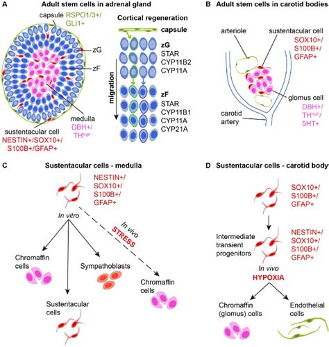
Chromaffin cells are the specialized neuroendocrine cells that constitute the bulk of the adrenal medulla. They are so named because of their characteristic staining properties with chromium salts, which cause them to turn brown. These cells are modified postganglionic sympathetic neurons that have lost their axons and instead secrete their neurotransmitters directly into the bloodstream. This endocrine mode of secretion differentiates them from typical neurons that release neurotransmitters at synapses.
Structure and Morphology of Chromaffin Cells
Chromaffin cells are characterized by their distinctive granular cytoplasm. These granules contain the catecholamines, epinephrine and norepinephrine, along with other proteins, such as chromogranins and enkephalins. The precise composition of these granules varies depending on the specific cell and its location within the medulla. Electron microscopy reveals a characteristic dense core within the granules, responsible for the chromaffin reaction. The cells themselves are polygonal in shape and typically arranged in clusters or cords, surrounded by a rich vascular network that facilitates the rapid release of hormones into the circulation.
Biosynthesis and Storage of Catecholamines
The process of catecholamine biosynthesis within chromaffin cells is a complex, multi-step pathway. It begins with the uptake of tyrosine from the bloodstream, which is then converted to L-DOPA by the enzyme tyrosine hydroxylase. This is the rate-limiting step in the pathway, subject to tight regulation. L-DOPA is subsequently converted to dopamine, then norepinephrine, and finally to epinephrine in those cells expressing the enzyme phenylethanolamine-N-methyltransferase (PNMT).
The synthesis of epinephrine distinguishes adrenal chromaffin cells from sympathetic neurons, which primarily produce norepinephrine. The stored catecholamines are packaged within the dense-core secretory granules, awaiting release upon stimulation. The precise control of this synthesis and storage ensures the availability of these crucial hormones when needed.
Release of Catecholamines: Mechanisms and Regulation
The release of catecholamines from chromaffin cells is triggered by the activation of nicotinic acetylcholine receptors on the cell surface. Upon binding of acetylcholine, released from preganglionic sympathetic neurons, these receptors initiate a cascade of intracellular events leading to exocytosis – the fusion of the secretory granules with the cell membrane and subsequent release of their contents into the extracellular space. Calcium ions play a critical role in this process, acting as a crucial intracellular messenger.
The release is also regulated by various other factors, including:
- Stress Hormones: Cortisol, released from the adrenal cortex, enhances the expression of PNMT, thus promoting epinephrine synthesis.
- Neural Inputs: The frequency and pattern of preganglionic sympathetic stimulation influence the amount and pattern of catecholamine release.
- Autocrine and Paracrine Factors: Local factors produced by chromaffin cells themselves, or by neighboring cells, can modulate catecholamine secretion.
The Developmental Origins of Chromaffin Cells: A Neural Crest Lineage
Chromaffin cells share a fascinating developmental origin with other components of the sympathetic nervous system: the neural crest. During embryonic development, neural crest cells migrate from the neural tube to various locations throughout the body, giving rise to a diverse array of cell types, including neurons, glial cells, and chromaffin cells. The specific signals and pathways that guide these neural crest cells to differentiate into chromaffin cells are an area of ongoing research. However, understanding this developmental origin highlights the close relationship between the adrenal medulla and the sympathetic nervous system.
Clinical Significance of Chromaffin Cell Dysfunction
Dysfunction of chromaffin cells can lead to a variety of clinical conditions, highlighting the importance of their normal function. Some key examples include:
Pheochromocytoma
Pheochromocytoma is a rare tumor arising from chromaffin cells. These tumors can produce excessive amounts of catecholamines, resulting in a constellation of symptoms, including hypertension, palpitations, headaches, and sweating. The diagnosis often involves measuring catecholamine levels in the urine or blood. Treatment strategies typically involve surgical removal of the tumor.
Neuroblastoma
Neuroblastoma is a cancerous tumor that originates from immature neural crest cells, which can differentiate into chromaffin-like cells. This cancer is most common in infants and young children. Diagnosis and treatment depend on the stage and characteristics of the tumor, and may involve surgery, chemotherapy, and radiation.
Research and Future Directions
Ongoing research continues to unravel the complexities of chromaffin cell function and its relevance to human health. Areas of focus include:
- Detailed understanding of the molecular mechanisms regulating catecholamine biosynthesis and release. This will aid in developing targeted therapies for conditions associated with chromaffin cell dysfunction.
- Exploration of the role of chromaffin cells in other physiological processes. While their role in stress response is well-established, their involvement in other pathways is gradually being understood.
- Development of novel diagnostic tools for detecting and monitoring chromaffin cell-related diseases. This is crucial for timely intervention and improving patient outcomes.
Conclusion: Chromaffin Cells – Essential Mediators of the Fight-or-Flight Response
The adrenal medulla, with its specialized chromaffin cells, stands as a crucial component of the body's stress response system. These cells, with their unique structure, biosynthesis pathways, and release mechanisms, are essential for the rapid production and release of epinephrine and norepinephrine into the circulation. Understanding the intricate details of chromaffin cell biology is critical, not only for appreciating the body's normal physiological responses but also for developing effective strategies to diagnose and treat conditions arising from their dysfunction. Further research into these fascinating cells promises to reveal even more about their intricate workings and broader impact on human health. Their study underscores the importance of interdisciplinary research combining cell biology, neuroendocrinology, and clinical medicine. The continued exploration of chromaffin cells will undoubtedly lead to further advancements in our understanding of the complex interplay between the nervous and endocrine systems, paving the way for improved diagnosis and treatment of various diseases.
Latest Posts
Latest Posts
-
Area Of A Parallelogram Cross Product
Apr 01, 2025
-
Completa Estas Oraciones Con Las Preposiciones Por O Para
Apr 01, 2025
-
Cartilage Is Separated From Surrounding Tissues By A Fibrous
Apr 01, 2025
-
Archaea And Bacteria Are Most Similar In Terms Of Their
Apr 01, 2025
-
Elements Are Organized On The Periodic Table According To
Apr 01, 2025
Related Post
Thank you for visiting our website which covers about What Secretory Cell Type Is Found In The Adrenal Medulla . We hope the information provided has been useful to you. Feel free to contact us if you have any questions or need further assistance. See you next time and don't miss to bookmark.
