What Vessels Connect To The Kidney In A Rat
Muz Play
Mar 15, 2025 · 7 min read
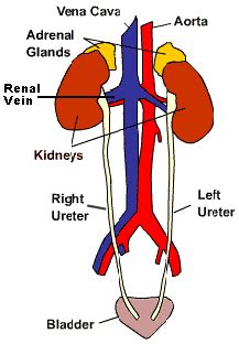
Table of Contents
What Vessels Connect to the Kidney in a Rat? A Comprehensive Overview
The rat, a common model organism in biomedical research, possesses a renal vascular system analogous to that of humans, yet with crucial anatomical differences. Understanding the precise vascular connections to the rat kidney is fundamental for researchers conducting studies on renal physiology, pharmacology, and pathology. This article provides a detailed exploration of the vessels connecting to the rat kidney, encompassing their origin, branching patterns, and functional significance.
Major Vessels Serving the Rat Kidney
The rat kidney's blood supply originates primarily from the renal artery, a branch of the abdominal aorta. This artery enters the kidney at the hilum, the indented region where the ureter, renal vein, and lymphatic vessels also enter and exit. The renal artery doesn't directly perfuse the nephrons; instead, it undergoes a complex series of branching events.
1. Renal Artery: The Primary Supply Line
The renal artery, upon entering the hilum, divides into several segmental arteries. The number of segmental arteries varies, but typically, a rat kidney will receive between two and four. This initial branching is crucial for distributing blood flow to different regions of the kidney. The precise number and arrangement can exhibit inter-individual variability, even within the same strain of rat. This inherent variability highlights the importance of careful anatomical observation in any experimental design involving renal vascular manipulations.
2. Interlobar Arteries: Delving Deeper
The segmental arteries further subdivide into interlobar arteries. These arteries travel within the renal columns, the cortical tissue extending between the renal pyramids. Their course is relatively straight, running towards the corticomedullary junction. The interlobar arteries are responsible for carrying oxygenated blood towards the nephrons located in the renal cortex and medulla. Their relatively straight trajectory minimizes resistance to blood flow, optimizing perfusion of the kidney parenchyma.
3. Arcuate Arteries: The Archway to Nephrons
At the corticomedullary junction, the interlobar arteries branch into arcuate arteries. These arteries form a characteristic arch-like structure along the base of the renal pyramids. The arcuate arteries are pivotal in supplying blood to both the cortex and the outer medulla. This strategic location ensures efficient oxygen and nutrient delivery to the nephrons situated in these regions, critical for their filtration and reabsorption functions. Disruption of the arcuate arteries can severely compromise kidney function.
4. Interlobular Arteries: Reaching the Nephrons
From the arcuate arteries, interlobular arteries arise. These vessels penetrate the renal cortex, branching repeatedly. They are responsible for delivering oxygenated blood directly to the glomeruli, the filtering units of the nephron. The interlobular arteries form a dense network within the cortex, ensuring thorough perfusion of the nephrons. Their intricate branching pattern allows for precise regulation of glomerular filtration rate (GFR), a crucial aspect of renal function. Studies investigating the effects of vasoactive agents often focus on the interlobular arteries due to their direct impact on GFR.
5. Afferent Arterioles: Gatekeepers of Filtration
The interlobular arteries ultimately give rise to afferent arterioles. These are the final branches before reaching the nephron. Each afferent arteriole supplies blood to a single glomerulus. The afferent arterioles are critically involved in regulating glomerular blood flow and thus GFR. Their diameter can be adjusted by the actions of various vasoactive substances, providing a key mechanism for maintaining blood pressure and fluid balance. The afferent arteriole's unique ability to constrict and dilate plays a crucial role in maintaining homeostasis.
The Venous Drainage System: Returning Blood to the Heart
The venous drainage of the rat kidney closely mirrors the arterial supply, albeit in reverse order. Blood, after passing through the nephrons and collecting the filtered waste products, is transported back to the heart via a series of interconnected veins.
1. Peritubular Capillaries and Vasa Recta: Collecting the Filtered Blood
The initial collection of deoxygenated blood occurs in the peritubular capillaries surrounding the proximal and distal convoluted tubules and the vasa recta, specialized capillaries surrounding the loops of Henle in the medulla. These capillaries are highly permeable, allowing for reabsorption of essential substances from the filtrate back into the bloodstream. The vasa recta, with their countercurrent exchange mechanism, are crucial for maintaining the medullary osmotic gradient, which is essential for concentrating urine.
2. Interlobular Veins: Convergence in the Cortex
The peritubular capillaries and vasa recta converge into interlobular veins. These veins collect blood from multiple nephrons and carry it towards the corticomedullary junction. They run parallel to the interlobular arteries, forming a dense network within the renal cortex. The interlobular veins play a critical role in transporting the blood back to the larger veins.
3. Arcuate Veins: Arching towards the Hilum
The interlobular veins coalesce to form arcuate veins, which, as their name suggests, form arches along the base of the renal pyramids. These veins run parallel to the arcuate arteries, mirroring their anatomical arrangement. The arcuate veins collect the blood from several interlobular veins and channel it toward the renal hilum.
4. Interlobar Veins: The Pathway to the Renal Vein
The arcuate veins converge into interlobar veins, which travel within the renal columns, towards the renal hilum. These veins run parallel to the interlobar arteries. The interlobar veins are essential for transporting the blood toward the main renal vein.
5. Renal Vein: Exit Point from the Kidney
Finally, the interlobar veins unite to form the renal vein. This large vein emerges from the hilum and carries the deoxygenated blood from the kidney back to the inferior vena cava, ultimately returning it to the heart. The renal vein's size reflects the substantial blood flow through the rat kidney.
Lymphatic Drainage of the Rat Kidney
Besides the arterial supply and venous drainage, the rat kidney also has a lymphatic system. Lymphatic vessels originating within the kidney drain into regional lymph nodes, contributing to the overall immune surveillance of the organ. These lymphatic vessels run alongside the renal artery and vein, draining lymph from the cortex and medulla. The lymphatic system plays a crucial role in removing waste products and immune cells from the kidney. Disruptions to the lymphatic drainage can lead to edema and impaired immune response within the kidney.
Functional Significance and Research Applications
The intricate vascular network of the rat kidney is not merely a passive conduit for blood; it's actively involved in regulating renal function. Understanding this network is crucial for:
-
Renal Physiology Studies: Researchers investigate the effects of various substances on glomerular filtration rate, renal blood flow, and tubular reabsorption using the rat as a model. Knowledge of the vascular anatomy is essential for precise targeting of experimental interventions.
-
Pharmacology Studies: The impact of new drugs on the kidney's vascular system is often assessed using rats. This involves investigating changes in renal blood flow, vascular resistance, and glomerular filtration in response to drug administration. Accurate knowledge of vascular anatomy ensures the correct interpretation of experimental results.
-
Pathology Studies: Rat models of kidney diseases, such as hypertension and diabetes, are widely used to study the pathogenesis and progression of renal damage. Understanding the vascular anatomy is critical for evaluating changes in blood flow and vascular structure associated with these diseases.
-
Surgical Procedures: Detailed knowledge of the renal vasculature is essential for planning and executing precise surgical procedures in the rat kidney, such as renal artery ligation or transplantation.
Conclusion: A Detailed Map for Renal Research
The intricate vascular network of the rat kidney, comprising the renal artery, its branching arteries, the venous system, and the lymphatic drainage, is a critical component of renal function. A thorough understanding of this vascular architecture is paramount for researchers investigating renal physiology, pharmacology, and pathology. This detailed overview serves as a comprehensive guide for researchers working with rat kidney models, facilitating precise experimental design and accurate interpretation of results. Further research continuously refines our understanding of the subtle variations and individual differences within this complex system, ensuring the accuracy and reproducibility of studies focused on the rat kidney.
Latest Posts
Latest Posts
-
Acids And Bases Cannot Mix Together
Mar 15, 2025
-
Linear Programming Do Not Find Minimum Or Maximum
Mar 15, 2025
-
The Change Rate Of Angular Momentum Equals To
Mar 15, 2025
-
Difference Between Tlc And Column Chromatography
Mar 15, 2025
-
Energy Required To Remove An Electron From A Gaseous Atom
Mar 15, 2025
Related Post
Thank you for visiting our website which covers about What Vessels Connect To The Kidney In A Rat . We hope the information provided has been useful to you. Feel free to contact us if you have any questions or need further assistance. See you next time and don't miss to bookmark.
