Identify The Body Cavities In The Following Illustration
Muz Play
Apr 01, 2025 · 7 min read
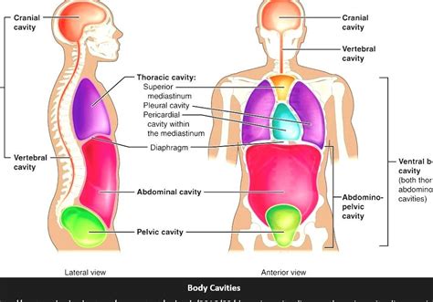
Table of Contents
Identifying Body Cavities: A Comprehensive Guide with Illustrations
Understanding the body's organizational structure is crucial in various fields, including medicine, anatomy, and physiology. A key aspect of this understanding involves recognizing the various body cavities, their locations, and the organs they house. This article provides a comprehensive overview of body cavities, using illustrative examples to clarify their positions and relationships. We will delve into the key cavities, their subdivisions, and the significance of their protective functions.
The Importance of Body Cavities
Body cavities serve several vital functions:
- Protection: They shield delicate organs from external shocks and impacts. Imagine the impact of a fall without the rib cage protecting your heart and lungs!
- Separation: Cavities allow for independent movement and function of different organ systems. This prevents interference between adjacent organs.
- Support: The skeletal framework provides structural support for the cavities, maintaining their shape and position.
- Compartmentalization: This allows for the independent functioning of organs within specific environments, including specialized temperature, pH, and fluid balance.
Major Body Cavities: A Detailed Exploration
The human body is broadly divided into two main cavities: the dorsal cavity and the ventral cavity. Let's explore these in detail:
1. Dorsal Cavity: Protecting the Central Nervous System
The dorsal cavity, located on the posterior side of the body, is subdivided into two smaller cavities:
1.1 Cranial Cavity: The Brain's Protective Shell
The cranial cavity is the superior part of the dorsal cavity, housed within the bony skull. It securely encloses and protects the brain, a vital organ controlling nearly all bodily functions. The cranial cavity's rigid bony structure provides excellent protection against trauma. The cerebrospinal fluid within the cavity further cushions the brain from impacts.
1.2 Vertebral (Spinal) Cavity: Protecting the Spinal Cord
Inferior to the cranial cavity lies the vertebral cavity, also known as the spinal cavity. This cavity runs along the length of the vertebral column (spine), protecting the delicate spinal cord. Similar to the cranial cavity, the vertebral cavity is surrounded by bone and contains cerebrospinal fluid to provide cushioning and protection. The spinal cord, a crucial component of the central nervous system, transmits signals between the brain and the rest of the body. Damage to the spinal cord within the vertebral cavity can lead to severe neurological impairments.
2. Ventral Cavity: Housing Vital Organs
The ventral cavity, situated on the anterior side of the body, is much larger than the dorsal cavity and contains the majority of the internal organs. It is further divided into the thoracic cavity and the abdominopelvic cavity.
2.1 Thoracic Cavity: A Complex Arrangement of Organs
The thoracic cavity, also known as the chest cavity, is superior to the abdominopelvic cavity and is separated from it by the diaphragm, a dome-shaped muscle crucial for respiration. The thoracic cavity is further divided into three smaller spaces:
-
Pleural Cavities (two): Each lung is enclosed within its own pleural cavity, a potential space lined by a serous membrane called the pleura. The pleura reduces friction during lung expansion and contraction. The pleural cavities help protect the lungs from injury and infection. Inflammation of the pleura (pleuritis) can cause significant pain.
-
Pericardial Cavity: This cavity is located within the mediastinum, a central region of the thoracic cavity that separates the lungs. The pericardial cavity surrounds the heart, providing protection and minimizing friction as the heart beats. The heart is also enclosed by a serous membrane called the pericardium. Fluid within the pericardial cavity cushions the heart and reduces friction.
-
Mediastinum: While not a cavity in the same sense as the pleural and pericardial cavities, the mediastinum is an important region within the thoracic cavity. It houses the heart, great blood vessels (aorta, vena cava, pulmonary arteries and veins), trachea, esophagus, and thymus gland.
2.2 Abdominopelvic Cavity: A Vast Space Containing Many Organs
The abdominopelvic cavity is the inferior portion of the ventral cavity, extending from the diaphragm to the pelvic floor. It is further subdivided into two smaller cavities: the abdominal cavity and the pelvic cavity. No physical structure completely separates these two, but the differences in their contents and location justify the subdivision.
-
Abdominal Cavity: The abdominal cavity contains most of the digestive organs, including the stomach, intestines (small and large), liver, gallbladder, pancreas, spleen, and kidneys. The abdominal cavity is lined by a serous membrane called the peritoneum, which helps protect the organs and reduces friction during movement. The peritoneum also forms mesenteries, supporting and suspending the abdominal organs.
-
Pelvic Cavity: The pelvic cavity is the inferior portion of the abdominopelvic cavity, located within the bony pelvis. It houses the urinary bladder, reproductive organs (uterus, ovaries, fallopian tubes in females; prostate gland, seminal vesicles in males), and the rectum. The pelvic cavity is also lined by the peritoneum.
Body Cavity Membranes: A Protective Lining
Serous membranes line the body cavities and cover the organs within them. These membranes secrete a lubricating fluid that reduces friction between the organs and the cavity walls. The serous membranes are crucial for preventing damage during organ movement. Each serous membrane consists of two layers:
- Parietal Layer: The outer layer lining the body cavity wall.
- Visceral Layer: The inner layer covering the organ.
The space between these two layers is called the serous cavity and contains a small amount of serous fluid. Examples include the pleura (lungs), pericardium (heart), and peritoneum (abdominal organs).
Clinical Significance of Body Cavities
Understanding the body cavities is essential for medical professionals for several reasons:
- Diagnosis: Pain or discomfort localized to a specific cavity helps pinpoint the possible source of the problem.
- Surgery: Knowledge of cavity boundaries and organ locations is crucial for safe and effective surgical procedures.
- Imaging: Medical imaging techniques like X-rays, CT scans, and MRI scans rely on understanding the location of the body cavities to interpret the images accurately.
- Trauma Treatment: Assessment of injuries affecting body cavities is critical for providing appropriate emergency care.
Abdominopelvic Regions and Quadrants: A Closer Look
To further enhance the localization of organs and pathologies, the abdominopelvic cavity is often divided into nine regions or four quadrants.
Nine Abdominopelvic Regions
These regions provide a more precise way to describe the location of organs and pain:
- Right Hypochondriac Region: Located under the ribs on the right side. Contains the liver, gallbladder, and parts of the intestines.
- Epigastric Region: Located in the upper central region, above the stomach. Contains the stomach, liver, and pancreas.
- Left Hypochondriac Region: Located under the ribs on the left side. Contains the spleen, stomach, and parts of the intestines.
- Right Lumbar Region: Located on the right side, between the hypochondriac and iliac regions. Contains parts of the intestines and the kidney.
- Umbilical Region: Located around the navel (umbilicus). Contains the transverse colon and small intestines.
- Left Lumbar Region: Located on the left side, between the hypochondriac and iliac regions. Contains parts of the intestines and the kidney.
- Right Iliac (Inguinal) Region: Located in the lower right region of the abdomen. Contains parts of the intestines, the appendix (often), and the female reproductive organs.
- Hypogastric (Pubic) Region: Located in the lower central region, above the pubic bone. Contains the urinary bladder, rectum, and reproductive organs.
- Left Iliac (Inguinal) Region: Located in the lower left region of the abdomen. Contains parts of the intestines and the female reproductive organs.
Four Abdominopelvic Quadrants
This simpler division uses two imaginary lines intersecting at the navel:
- Right Upper Quadrant (RUQ): Contains the liver, gallbladder, parts of the intestines, and the right kidney.
- Left Upper Quadrant (LUQ): Contains the stomach, spleen, parts of the intestines, and the left kidney.
- Right Lower Quadrant (RLQ): Contains the appendix, parts of the intestines, and the female reproductive organs.
- Left Lower Quadrant (LLQ): Contains parts of the intestines and the female reproductive organs.
Understanding the body cavities and their subdivisions is crucial for comprehending human anatomy and physiology. This knowledge forms the foundation for diagnosis, treatment, and research in various medical fields. While this article provides a comprehensive overview, further exploration of specific organs and their relationships within these cavities will enrich your understanding of the human body's intricate design. Remember to consult reliable anatomical resources and texts for a more detailed and nuanced understanding.
Latest Posts
Latest Posts
-
Confidence Interval Calculator For 2 Proportions
Apr 02, 2025
-
Draw A Structural Formula For 3 Bromo 4 Chloro 1 1 Dimethylcyclohexane
Apr 02, 2025
-
What Is A Primary Standard Chemistry
Apr 02, 2025
-
How Do You Convert From Atoms To Grams
Apr 02, 2025
-
How Do Positive Ions And Negative Ions Form
Apr 02, 2025
Related Post
Thank you for visiting our website which covers about Identify The Body Cavities In The Following Illustration . We hope the information provided has been useful to you. Feel free to contact us if you have any questions or need further assistance. See you next time and don't miss to bookmark.
