The Neural Tunic Of The Eye
Muz Play
Mar 28, 2025 · 7 min read
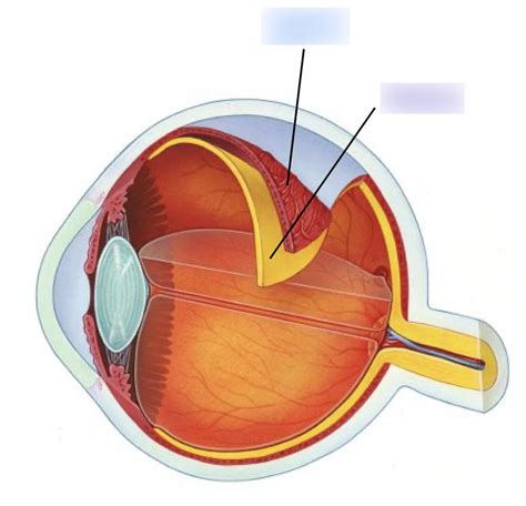
Table of Contents
The Neural Tunic of the Eye: A Deep Dive into the Retina and its Crucial Role in Vision
The eye, a marvel of biological engineering, allows us to perceive the world around us in stunning detail. At the heart of this visual experience lies the neural tunic, more commonly known as the retina. This complex, multilayered structure is responsible for converting light into electrical signals that the brain can interpret as images. Understanding its intricate anatomy, physiology, and potential pathologies is crucial for appreciating the complexities of vision and diagnosing related disorders.
The Retina: A Multilayered Masterpiece
The retina, a thin, delicate membrane lining the inner surface of the eye, is not a single uniform layer but rather a sophisticated assembly of distinct cell types, each playing a vital role in visual processing. These layers are intricately connected, facilitating the rapid and efficient transformation of light into neural signals.
Key Cellular Components of the Retina:
-
Photoreceptor Cells: These are the primary light-sensitive cells, responsible for capturing photons and initiating the visual transduction cascade. There are two main types:
- Rods: Highly sensitive to light, enabling vision in low-light conditions. They are responsible for scotopic vision (night vision) and lack the ability to distinguish colors.
- Cones: Less sensitive to light but crucial for color vision and visual acuity. They function optimally in bright light conditions (photopic vision) and are concentrated in the macula, especially the fovea.
-
Bipolar Cells: These cells act as intermediaries, receiving signals from photoreceptors and transmitting them to ganglion cells. They play a critical role in shaping the visual signal, enhancing contrast and sensitivity.
-
Ganglion Cells: These are the output neurons of the retina, their axons forming the optic nerve, which carries visual information to the brain. Different types of ganglion cells contribute to various aspects of vision, such as motion detection, contrast sensitivity, and color perception.
-
Horizontal Cells: These cells form lateral connections between photoreceptors and bipolar cells, mediating lateral inhibition. This process enhances contrast and edge detection, sharpening visual images.
-
Amacrine Cells: These cells also contribute to lateral interactions, mainly influencing the signals between bipolar and ganglion cells. They play a vital role in adapting to different light levels and contributing to various aspects of visual processing, including motion detection and temporal resolution.
-
Müller Cells: These are glial cells that provide structural support and metabolic function to the retina. They also play a critical role in maintaining the ionic balance and removing waste products.
The Retinal Circuitry: A Symphony of Signals
The interaction between these retinal cell types creates a complex neural circuit that processes visual information with remarkable efficiency. The process begins when light photons strike the photoreceptors. This triggers a cascade of biochemical events, ultimately leading to a change in the membrane potential of the photoreceptor cells.
This change in membrane potential is then relayed through the bipolar cells to the ganglion cells. Horizontal and amacrine cells modulate this signal transmission, refining the information and enhancing its contrast and sharpness. The signals from the ganglion cells are then transmitted along the optic nerve to the brain's visual cortex for final interpretation.
Visual Transduction: The Conversion of Light into Electrical Signals
Visual transduction, the process of converting light energy into electrical signals, is a complex multi-step process within the photoreceptor cells. This process begins with the absorption of photons by photopigments, specialized proteins embedded in the membranes of the photoreceptors.
In rods, the photopigment is rhodopsin, while cones contain different types of photopigments (opsins) that are sensitive to different wavelengths of light, enabling color vision. Absorption of a photon leads to a conformational change in the photopigment, initiating a cascade of biochemical reactions that ultimately result in a hyperpolarization of the photoreceptor cell membrane.
This hyperpolarization reduces the release of neurotransmitters, thereby altering the activity of the downstream bipolar and ganglion cells. This change in neurotransmitter release is the crucial step in translating light into a neural signal.
The Macula and Fovea: Regions of High Acuity
The macula is a small, specialized region of the retina located near the center of the visual field. It is responsible for sharp, detailed central vision. Within the macula lies the fovea, a tiny pit containing a high concentration of cones and almost no rods. This anatomical arrangement accounts for the fovea's exceptional visual acuity.
The absence of other retinal layers overlying the photoreceptors in the fovea allows light to reach the cones directly, minimizing scattering and maximizing the clarity of the image. This ensures the highest level of visual resolution, critical for tasks requiring fine detail, such as reading or recognizing faces.
Clinical Relevance: Diseases and Disorders of the Neural Tunic
The retina's vital role in vision makes it susceptible to various diseases and disorders. These can significantly impact visual function, potentially leading to blindness if left untreated.
Common Retinal Pathologies:
-
Age-Related Macular Degeneration (AMD): A leading cause of vision loss in older adults, AMD involves the progressive deterioration of the macula, leading to blurred central vision and ultimately blindness.
-
Diabetic Retinopathy: A complication of diabetes, this condition damages the blood vessels in the retina, leading to blurred vision, floaters, and potentially blindness.
-
Retinitis Pigmentosa: A group of inherited retinal diseases characterized by progressive degeneration of photoreceptors, resulting in night blindness and a gradual loss of peripheral vision.
-
Retinal Detachment: A condition where the retina separates from the underlying choroid, causing a loss of vision. Prompt treatment is essential to prevent permanent vision loss.
-
Glaucoma: While not directly a retinal disease, glaucoma damages the optic nerve, often leading to characteristic changes in the retinal nerve fiber layer. Early detection and management are crucial to prevent irreversible vision loss.
Advanced Imaging Techniques for Retinal Assessment
Modern ophthalmology employs advanced imaging techniques to visualize and assess the health of the retina in unprecedented detail. These techniques allow for early detection and monitoring of retinal diseases, improving patient outcomes.
-
Optical Coherence Tomography (OCT): This non-invasive imaging technique provides high-resolution cross-sectional images of the retina, allowing for detailed visualization of its layers and identification of subtle structural changes indicative of disease.
-
Fluorescein Angiography (FA): This technique uses a fluorescent dye to visualize the retinal blood vessels, revealing abnormalities such as leakage or blockage that may be indicative of various retinal pathologies.
-
Fundus Photography: This technique captures high-resolution images of the fundus (the back of the eye), allowing for documentation of retinal features and changes over time.
Future Directions in Retinal Research
Research into the retina and its related disorders is constantly evolving, driven by the desire to develop innovative therapies and improve the lives of individuals affected by retinal diseases. Current research focuses on:
-
Gene therapy: This approach aims to correct genetic defects responsible for inherited retinal diseases, offering the potential for restoring vision.
-
Stem cell therapy: This promising area of research explores the use of stem cells to replace damaged photoreceptors or other retinal cells.
-
Pharmacological interventions: Research continues to identify and develop new drugs to treat various retinal diseases, slowing progression and preserving vision.
-
Neuroprosthetics: This emerging field explores the development of artificial retinas or other neuroprosthetic devices to restore vision in individuals with severe retinal damage.
Conclusion: The Retina—A Vital Component of Vision
The neural tunic of the eye, the retina, is a marvel of biological engineering, a complex and delicately balanced structure responsible for transforming light into the images we perceive. Its intricate cellular organization, sophisticated circuitry, and susceptibility to various diseases make it a fascinating and crucial area of study. Ongoing research continues to unlock the secrets of the retina, paving the way for improved diagnosis, treatment, and ultimately, the preservation of sight for countless individuals. Understanding the complexity and importance of the retina reinforces the significance of regular eye examinations and the ongoing pursuit of advancements in retinal research.
Latest Posts
Latest Posts
-
Why Is Mol The Abbreviation To Mle
Mar 31, 2025
-
What Is The Correct General Equation For Cellular Respiration
Mar 31, 2025
-
Which Element Is The Least Reactive
Mar 31, 2025
-
Which One Is Good Insulator Metals Metalloids Or Nonmetals
Mar 31, 2025
-
Why Are Covalent Compounds Not Conductive
Mar 31, 2025
Related Post
Thank you for visiting our website which covers about The Neural Tunic Of The Eye . We hope the information provided has been useful to you. Feel free to contact us if you have any questions or need further assistance. See you next time and don't miss to bookmark.
