What Color Is An Animal Cell
Muz Play
Mar 26, 2025 · 5 min read
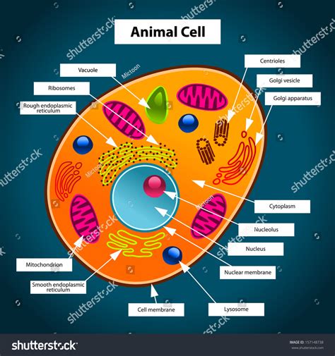
Table of Contents
What Color Is an Animal Cell? Exploring the Nuances of Cellular Visibility
The question, "What color is an animal cell?" might seem deceptively simple. However, the answer is far more nuanced than a single color. The reality is that animal cells, in their natural state, are largely colorless or translucent. Their appearance depends heavily on several factors, including the type of cell, staining techniques used for observation, and the magnification of the microscope employed. This article delves deep into the complexities of visualizing animal cells and explores the various factors influencing their perceived color.
The Inherent Transparency of Animal Cells
Unlike some plant cells, which contain chlorophyll and appear green, animal cells lack pigments that impart a distinct hue. Their cytoplasm, the jelly-like substance filling the cell, is primarily composed of water, proteins, and other colorless molecules. This lack of intrinsic coloration makes them virtually invisible to the naked eye. Their translucent nature is a crucial aspect of their functionality, allowing light to pass through them, which is essential for various cellular processes.
The Role of Organelles
While the overall cell is generally colorless, the internal structures, or organelles, within the cell can have subtle variations in light refraction. However, these differences are typically too minute to be observed without specialized techniques. For instance, the nucleus, containing the cell's genetic material, might appear slightly denser and therefore less translucent than the surrounding cytoplasm, but this difference is not a distinct color.
Enhancing Visibility: The Importance of Staining
To visualize the intricate details of animal cells and their components, scientists employ staining techniques. These techniques use dyes that bind to specific cellular structures, making them readily visible under a microscope. The choice of stain directly influences the perceived color of the cell and its components.
Common Staining Techniques and Their Color Impacts
Several staining techniques are commonly used to visualize animal cells:
-
Hematoxylin and Eosin (H&E) staining: This is a widely used stain in histology (the study of tissues). Hematoxylin stains the cell nucleus a deep purple or blue, while eosin stains the cytoplasm a pinkish or reddish hue. This combination provides excellent contrast and allows for the identification of different cellular structures.
-
Wright's stain: Frequently used in hematology (the study of blood), Wright's stain is a differential stain, meaning it stains different cellular components with different colors. It's particularly useful for identifying various blood cell types, staining nuclei purple and cytoplasm shades ranging from light pink to deep blue-purple, depending on the cell type.
-
Gram staining: While primarily used for bacteria, Gram staining can also be applied to some animal cells, particularly those with a thick cell wall. Gram-positive cells typically stain purple, while Gram-negative cells appear pink. This staining method provides crucial information about cell wall structure and composition.
-
Immunofluorescence staining: This advanced technique uses fluorescent antibodies to target specific proteins or other molecules within the cell. These antibodies emit light of various colors (green, red, blue, yellow) when excited by specific wavelengths of light, allowing researchers to visualize the location and abundance of target molecules within the cell. This technique allows for the visualization of intricate cellular processes and interactions.
-
Phase-contrast microscopy: This technique doesn't involve staining but rather exploits the differences in refractive index between different parts of the cell. This allows for the visualization of cellular structures without the need for staining, resulting in a mostly colorless image, but with subtle variations in brightness and contrast revealing the internal structures.
The Impact of Microscope Type and Magnification
The perceived color of an animal cell is also heavily influenced by the type of microscope used and the magnification level. Different microscopes employ different mechanisms to visualize cells, leading to variations in contrast and color representation.
Light Microscopy
Light microscopy is the most commonly used method for visualizing animal cells. Depending on the staining technique used, the cell and its components might appear in various colors, as described in the previous section. However, even without staining, the resolution of light microscopy allows for the observation of subtle differences in light refraction, resulting in a gray-scale image with variations in brightness.
Electron Microscopy
Electron microscopy provides much higher resolution than light microscopy, allowing for the visualization of subcellular structures in far greater detail. However, electron microscopy doesn't directly provide colored images. The images produced are in grayscale, with different densities represented by various shades of gray. False color is often artificially added to electron micrographs to enhance the visualization and understanding of the structures.
Factors Affecting Apparent Color Beyond Staining
Beyond the deliberate use of stains, several other factors can influence the apparent color of an animal cell, even under the microscope:
-
Artifacts: During sample preparation, various artifacts might be introduced, leading to variations in appearance. These artifacts can be mistaken for actual cellular components, potentially leading to misinterpretations of the color and structure.
-
Background Interference: The surrounding medium and any debris or contamination in the sample can interfere with the visualization of the cell, altering its apparent color.
-
Optical Properties: Different microscope settings, such as lighting and filter usage, can influence the perceived color and brightness of the cell.
Conclusion: The Multifaceted Nature of Animal Cell Color
In conclusion, the question of what color an animal cell is does not have a simple, single answer. In their natural state, animal cells are largely colorless and translucent. However, the use of various staining techniques and microscopy methods allows for visualization and creates apparent colors. The perceived color depends on the staining method, the type of microscopy used, the magnification, and other factors such as artifacts and background interference. Therefore, the “color” of an animal cell is a multifaceted concept dependent on the specific experimental context and techniques employed. Understanding these complexities is crucial for accurate interpretation of cellular images and a deeper understanding of cell biology.
Latest Posts
Latest Posts
-
Properties Of Ionic And Covalent Compounds Lab
Mar 27, 2025
-
What Is The Electron Configuration Of B
Mar 27, 2025
-
What Is Periodic Motion In Physics
Mar 27, 2025
-
How To Factor Trinomials With Leading Coefficient
Mar 27, 2025
-
How Much Atp Is Produced In Oxidative Phosphorylation
Mar 27, 2025
Related Post
Thank you for visiting our website which covers about What Color Is An Animal Cell . We hope the information provided has been useful to you. Feel free to contact us if you have any questions or need further assistance. See you next time and don't miss to bookmark.
